In vitro assays for enriching and determining the clonogenic potential of stem cells

a technology of stem cell clone and in vitro assay, which is applied in the field of in vitro assays for enriching and determining the clone potential of stem cells, can solve the problems of not observing enhanced self-renewal or tumor initiating capacity, time-consuming process, and complex methods, and achieve the effect of reducing concerns about clone aggregation

Active Publication Date: 2013-01-10
UNIVERSITY OF ROCHESTER
View PDF1 Cites 4 Cited by
  • Summary
  • Abstract
  • Description
  • Claims
  • Application Information

AI Technical Summary

Benefits of technology

[0013]The accompanying Examples also demonstrate, in one embodiment, the use of PDMS microbubble arrays to determine the clonogenic potential of these enriched NA / S and AM sub-populations. Microbubbles are spherical cavities formed in PDMS using the gas expansion molding (GEM) process (Giang et al., “Microfabrication of Cavities in Polydimethylsiloxane using DRIE Silicon Molds,”Lab Chip 7(12):1660e2 (2007), which is hereby incorporated by reference in its entirety). The microbubble cell culture platform is ideally suited for studying CSCs in the tumor microenvironmental niche, which is composed of soluble factors, extracellular matrix (ECM) proteins, cell-cell contacts and mechanical loading. These factors determine the clonogenic and tumorigenic potential in vivo and govern whether a cell can self-renew and / or differentiate into a tumor or normal cell or remain quiescent (Bissell et al., “Context, Tissue Plasticity, and Cancer: Are Tumor Stem Cells also Regulated by the Microenvironment?”Cancer Cell 7(1):17e23 (2005); Ghotra et al., “The Cancer Stem Cell Microenvironment and Anti-cancer Therapy,”Int J Radiat Biol 85(11):955e62 (2009), each of which is hereby incorporated by reference in its entirety). The advantage of using microbubbles for culturing enriched cell sub-populations derives from the fact that all the properties of the cell microenvironment can be specifically investigated; the geometry allows for the accumulation of autocrine / paracrine factors while permitting sufficient nutrient and waste exchange with the bulk media reservoir through the microbubble opening (Chandrasekaran et al., “Microenvironment Induced Spheroid to Sheeting Transition of Immortalized Human Keratinocytes (HaCaT) Cultured in Microbubbles Formed in Polydimethylsiloxane,”Biomaterials 32(29):7159e68 (2011), which is hereby incorporated by reference in its entirety). The surface of microbubble can be coated with proteins of interest (Giang et al., “Microfabrication of Bubbular Cavities in PDMS for Cell Sorting and Microcell Culture Applications,”J Bionic Eng 5(4):308e16 (2008), which is hereby incorporated by reference in its entirety), there is room for cell-cell contacts inside the microbubble and the elastic modulus of PDMS closely resembles in vivo tissue thereby providing a more realistic mechanical load (Janmey and Miller, “Mechanisms of Mechanical Signaling in Development and Disease,”J Cell Sci 124(Pt 1):9e18 (2011), which is hereby incorporated by reference in its entirety). The microbubble cell culture platform enables rapid (˜5-7 days) generation of pure clones from single cells and eliminates concerns for clone aggregation that may occur when cells are plated at high clonal densities to enable their survival in standard limiting dilution assay conditions (Pastrana et al., “Eyes Wide Open: A Critical Review of Sphere-formation as an Assay for Stem Cells,”Cell Stem Cell 8(5):486e98 (2011); Fedoroff and Richardson, “Colony Cultures—Plating Efficiency Assay and Cloning, in Protocols for Neural Cell Culture,” 307e315, Humana Press, doi:10.1385 / 1-59259-207-4 (2001), each of which is hereby incorporated by reference in its entirety).

Problems solved by technology

They did not, however, observe enhanced self-renewal or tumor initiating capacity in xenotransplantation experiments.
These methods are complex, and expensive as growth is usually done under limiting dilution conditions where cells are plating at low density (≦1 cell per well).
This is a time consuming process (3-4 weeks), that often requires special media that must be developed through tedious trial-and-error processes.

Method used


Image

  • In vitro assays for enriching and determining the clonogenic potential of stem cells
  • In vitro assays for enriching and determining the clonogenic potential of stem cells
  • In vitro assays for enriching and determining the clonogenic potential of stem cells

Examples


example 1

Morphology of WM115 Cell Line on Planar PDMS and TCP

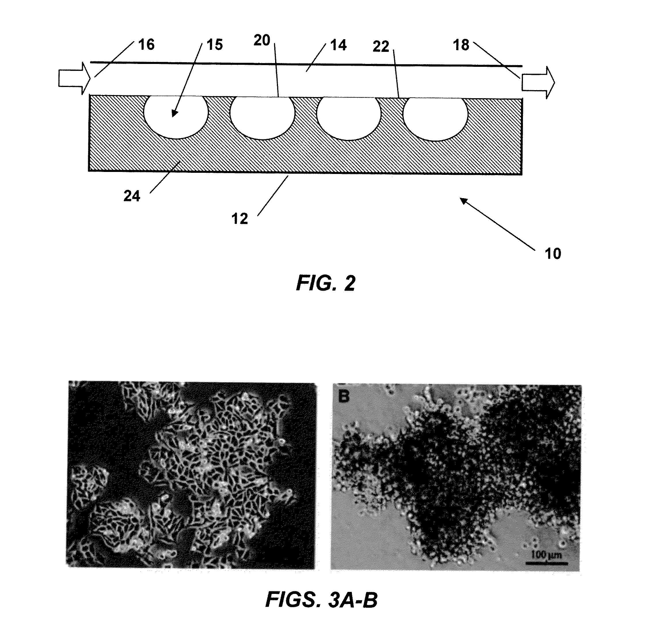
[0092]WM115 cells were seeded at a density of 2×10⁴ cells / cm2 on TCP and planar PDMS and cultured as described in the preceding Materials and Methods. Results indicated that WM115 cells propagate as a monolayer on TCP (FIG. 3A); however, on planar hydrophobic PDMS they tend to propagate with a non-adherent 3D cluster morphology (FIG. 3B). Plasma treated TCP is hydrophilic, which favors cell adhesion and spreading, whereas PDMS is a hydrophobic polymer that hinders cell adhesion causing cells to adopt 3D cluster morphology.

example 2

Viability of WM115 Cell Line on TCP and Planar PDMS

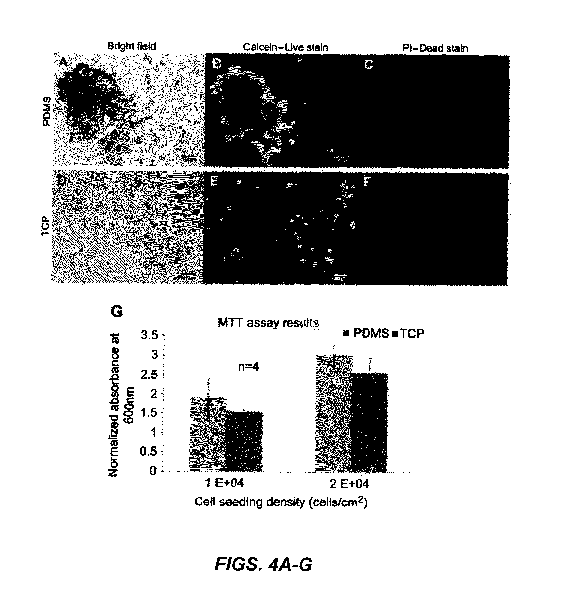
[0093]Cell viability was tested using an assay with calcein-AM and propidium iodide for live and dead stain, respectively. Calcein-AM is membrane permeable and is cleaved by active esterases in live cells which produce a fluorescence signal. Propidium iodide is cell membrane impermeable, however, dead cells that have degraded cell membranes allow the stain to enter where it intercalates DNA. The stains were applied to the cells after 4 days of incubation on planar PDMS and TCP (FIGS. 4A, 4D) as described in Section 2.4 and results showed that most cells were viable based on fluorescence visualization. Cells propagating as 3D clusters and monolayer on TCP stain positive for the live stain (FIGS. 4B, 4E) and there is very little staining for dead stain (FIGS. 4C, 4F). This suggested that there was sufficient nutrient uptake by cells propagating as 3D clusters on planar PDMS.

[0094]MTT assay results show the effect of the PDMS substrate...

example 3

Effect of Sub-Culturing WM115 Cell Line from Planar PDMS to TCP

[0095]WM115 cells propagating as 3D clusters on planar PDMS were non-adherent to the underlying substrate and pipetting the media ensured the removal of cells from the well. When the cells cultured on planar PDMS were harvested, dissociated and plated (seeding density=1×10⁴ cells / cm2) as individual cells on TCP (FIG. 5A), cells with two distinct morphology emerged on TCP by the end of day 4 (FIG. 5B). WM115 cells exhibited a unique “fried egg” morphology comprised of cells that formed tightly packed non-adherent spheroids (NA / S) in the center surrounded by cells that propagated as an adherent monolayer (AM).


PUM

PropertyMeasurementUnit
elastic modulusaaaaaaaaaa

Abstract

A method of enriching stem or progenitor cells that includes growing a heterogeneous cell sample comprising stem and/or progenitor cells on a first substrate that is hydrophobic and has an elastic modulus less than about 100 MPa; recovering the heterogeneous cell sample from the first substrate; growing the recovered heterogeneous cell sample on a second substrate that is hydrophilic and has an elastic modulus higher than the elastic modulus of the first substrate to produce a subpopulation of nonadherent cells and a subpopulation of adherent cells; and recovering the nonadherent cell subpopulation, which is enriched for stem and/or progenitor cells. The invention also relates to a method of determining the clonogenic potential of a cell, such as a cancer stem cell enriched using the enrichment method described herein.

Description

[0001]This application claims the priority benefit of U.S. Provisional Patent Application Ser. No. 61 / 505,881, filed Jul. 8, 2011, which is hereby incorporated by reference in its entirety.[0002]This invention was made with support from the National Science Foundation under grant CBET 0827862. The U.S. government has certain rights in this invention.FIELD OF THE INVENTION[0003]This invention relates to novel assays for the enrichment of stem cells (somatic or tumor initiating), and determining the clonogenic potential of such cells. These assays can also be used to sort tumor initiating cells, screen new drugs for their ability to destroy tumor initiating cells or retard their clonogenic potential. The assays are also useful for sorting and characterizing somatic stem or progenitor cells.BACKGROUND OF THE INVENTION[0004]Somatic (adult) stem cells maintain tissue homeostasis by exhibiting long-term replicative potential, together with the capacities of self-renewal and multi-lineage ...

Claims


Application Information

Patent Timeline
Patent Type & Authority Applications(United States)
IPC IPC(8): C40B30/06C12N5/071
CPCG01N33/5073C12N5/0695C12N2539/10C12M23/12C12N2533/30
Inventor DELOUISE, LISACHANDRASEKARAN, SIDDARTH
Owner UNIVERSITY OF ROCHESTER
Why Eureka
Try Eureka