
Direct protein delivery with engineered microvesicles

a technology of microvesicles and direct protein, which is applied in the direction of viruses/bacteriophages, antibody medical ingredients, peptide sources, etc., can solve the problems of limiting their use, undesirable effects, and slow expression of desired proteins

Active Publication Date: 2012-12-20
INST NAT DE LA SANTE & DE LA RECHERCHE MEDICALE (INSERM)
View PDF1 Cites 22 Cited by
  • Summary
  • Abstract
  • Description
  • Claims
  • Application Information

AI Technical Summary

Benefits of technology

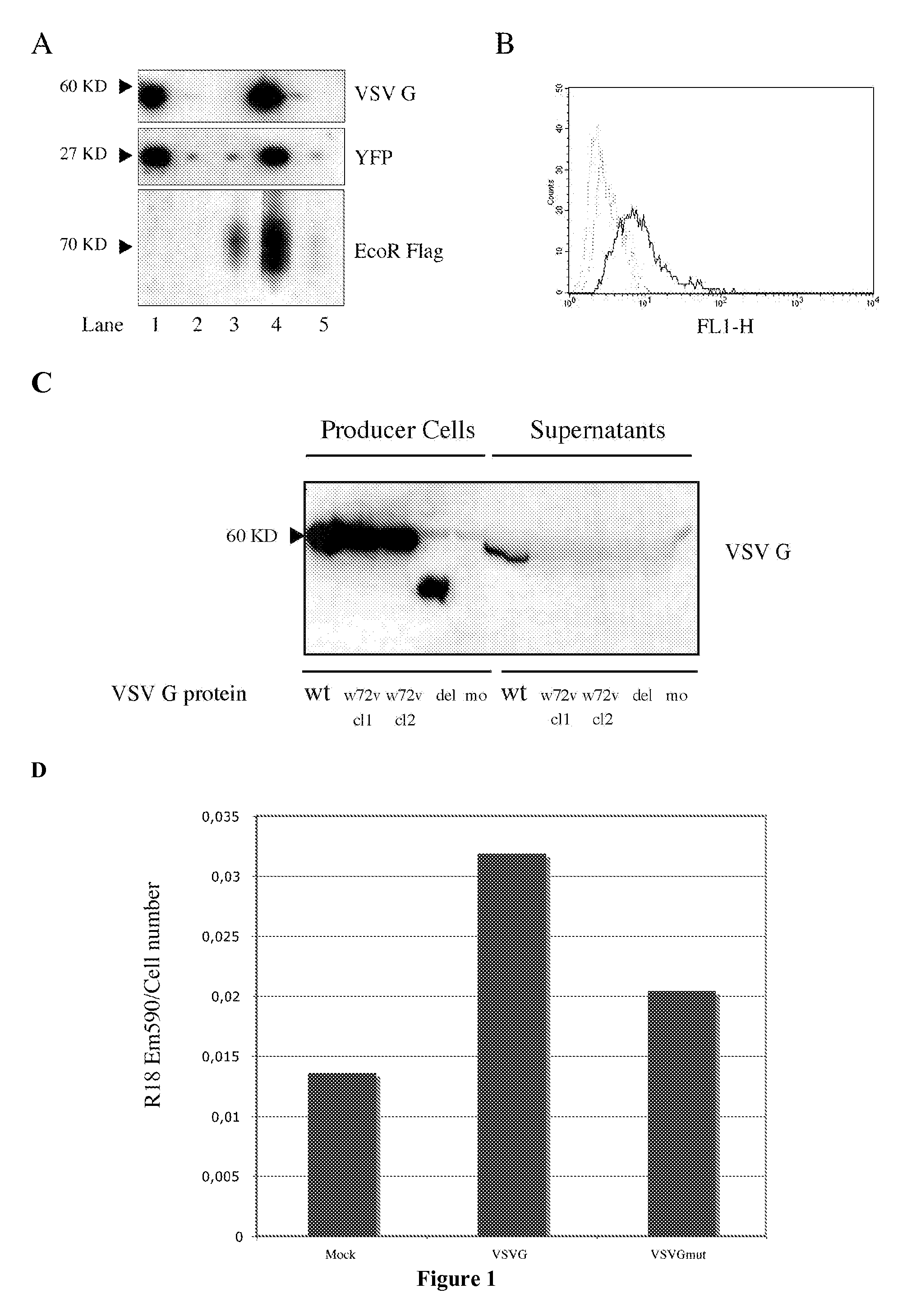
[0014]The inventors have discovered that the overexpression of a viral membrane fusion protein in a eukaryotic cell expressing a protein of interest can lead to the secretion of microvesicles comprising said membrane fusion protein and said protein of interest. Further, the inventors have demonstrated that said microvesicles can be used to efficiently deliver said protein of interest to a target cell, without altering the function of said protein. The method of the present invention is thus suitable for the transient and rapid introduction of a functional protein in a target cell.
[0079]Due to the low amounts of material delivered by the microvesicles according to the invention and to their non-genetic nature, the microvesicles appear to be useful for applications where low and transient presence of proteins may lead to striking biological effects, i.e. to proteins having a high biological penetrance.

Problems solved by technology

The transient introduction of proteins of interest into a target cell is a major challenge, with many applications in basic research, applied science and in the therapeutic field.
However, these techniques have several inconvenients that many limit their use.
The expression of the desired protein is thus slow, and several undesirable effects can be observed (toxicity of the transfection reagent itself, activation of the interferon response etc.).
Moreover, certain cell types, typically non-dividing cells such as neurons, are poorly transfectable.
However, these technologies suffer from several drawbacks.
First, there is a sanitary risk associated with the use of retroviral vector, even though said retroviral vectors have been inactivated.
Second, production of retroviral particles is very tedious and time-consuming.
However, this technique requires the obtaining of the protein of interest prior to microinjection.
Thus, these methods are costly and poorly amenable to large-scale uses.
However, these techniques are also difficult to implement on a large-scale basis, since they rely on recombinant proteins.

Method used


Image

  • Direct protein delivery with engineered microvesicles
  • Direct protein delivery with engineered microvesicles
  • Direct protein delivery with engineered microvesicles

Examples


example 1

[0165]Abstract

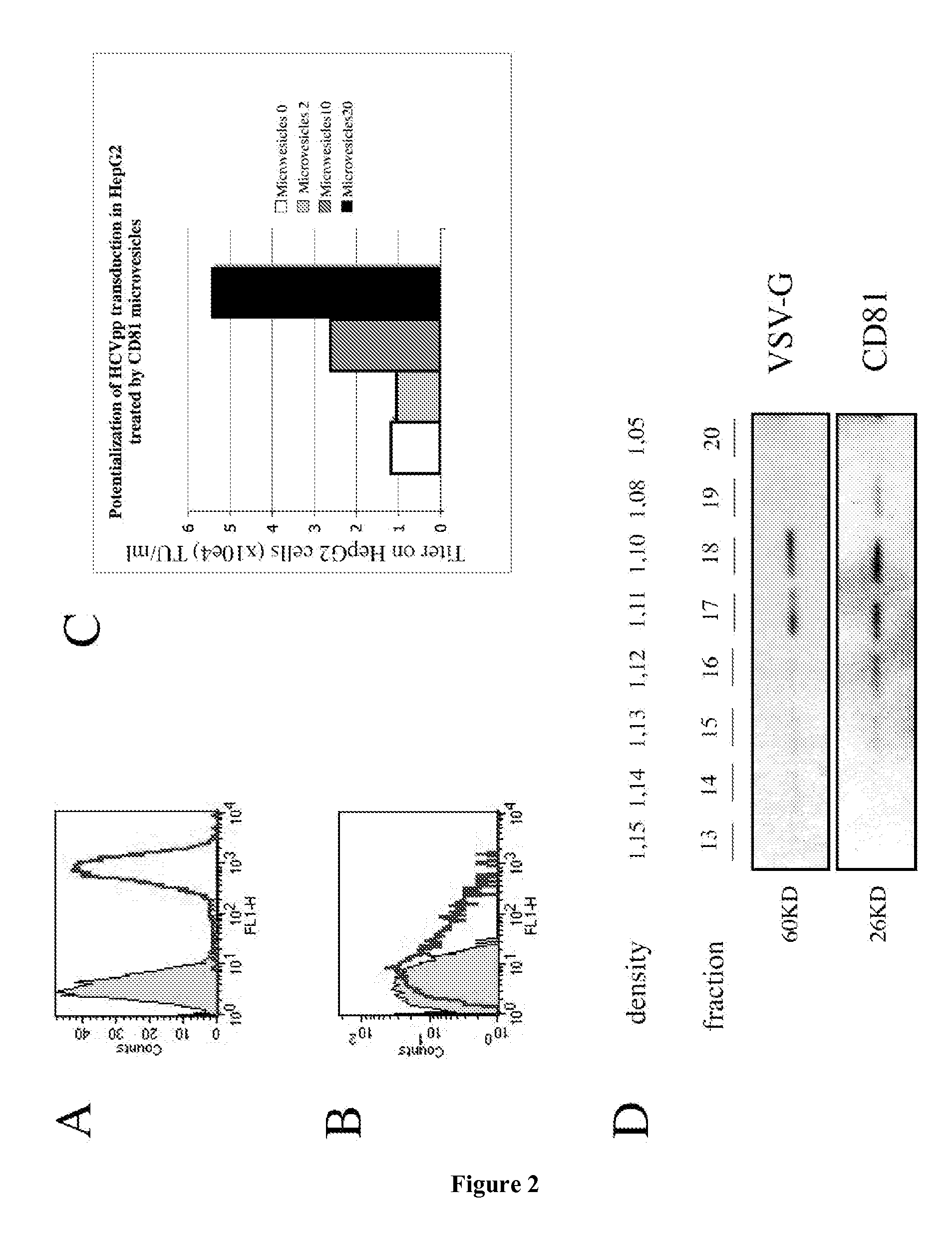
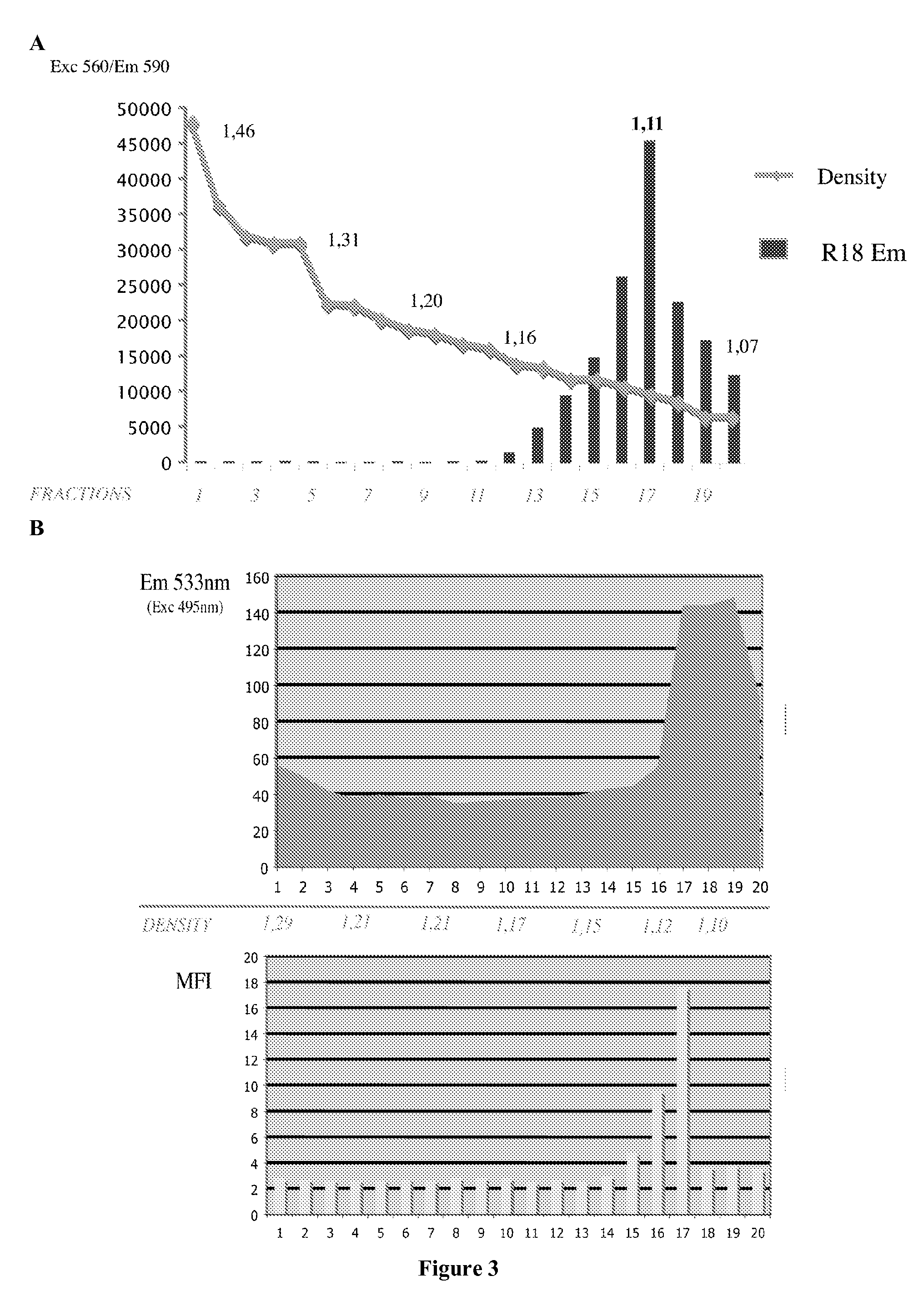
[0166]The present example describes the engineering of exosome-like vesicles coated with the G glycoprotein of the Vesicular Stomatitis Virus (VSV-G) and used to deliver exogenous proteins in human target cells. These particles sediment at a density of 1.1 g / ml and can efficiently package cytoplasmic and membrane proteins as well as transcription factors. Model molecules including the membrane mCAT-1 protein and the tet transactivator were efficiently delivered in human target cells upon microvesicles treatment. Vesicle-mediated pseudotransduction led to transient protein expression and was not due to transfer of mRNA or DNA. We showed that chloroquine, a drug raising endosomal pH, invalidated protein transfer indicating that VSV-G induced fusion was involved in the delivery mechanism.

[0167]The VSV-G Fusogenic Envelope is a Key Determinant of Pseudotransduction

[0168]We tested whether expression of VSV-G could lead to the production of particles able to pseudotransduce....

example 2

[0205]Vesicle-mediated protein transfer in a coculture experiment through 3-μm pore sized inserts.

[0206]We cultivated in the same media HEK cells transfected by various combinations of plasmids, together with the Teo-GFP reporter cell line. Cells were separated by a 3-μm pore sized filter as depicted, the producing cells in the bottom of a 6-well dish and the reporter cell line in the top insert. Producing cells were cotransfected with a tTA expression plasmid in addition with a carrier DNA, the VSV-G coding plasmid or a fusion defective VSV-G. After 48 hours of coculture, inserts were removed and the reporter cell line trypsinized and analysed by FACS to evaluate tTA delivery. Results are given as the MFI in the Teo-GFP cell line (FIG. 12).

[0207]This technique enables the delivery of protein into target cells, without the need of a concentration step. Concentrated vesicles can be toxic depending on the target cell type. Target cells can be cultivated in the top or the bottom chambe...

example 3

[0208]Insect Cells Microvesicle Production and Dosage

[0209]Baculovirus production was performed using the Bac-to-Bac Baculovirus expression system (Invitrogen) according to the manufacturer's instructions. Briefly, cDNAs of interest were cloned in the pFAST-1 shuttle prior to recombination in DH10-BAC bacteria. Baculovirus DNAs were next used to transfect SF-9 cells to generate the first baculo stock collected 72 hours later (passage 1). This polyclonal stock was further amplyfied to reach a titer of 1×10⁷ pfu / ml (passage 2) that was stored at 4° C. and used for vesicles production.

[0210]Insect cells microvesicles were produced upon baculovirus infection (MOI 0.5-1) of 200×106 HIGH5 cells cultivated in suspension at 30° C. under agitation in 100 ml of Express-five SFM media supplemented with Glutamine (20 mM) and Penicillin-Streptomycin (25U of penicillin, 25 μg of Streptomycin / nil). 48 hours after inoculation, medium was harvested clarified and filtered twice through a 0.45 μm pore...


PUM

PropertyMeasurementUnit
sizeaaaaaaaaaa
sizeaaaaaaaaaa
sizeaaaaaaaaaa

Abstract

The present invention relates to direct protein delivery with engineered micro vesicles.

Description

FIELD OF THE INVENTION[0001]The present invention relates to direct protein delivery with engineered microvesicles.BACKGROUND OF THE INVENTION[0002]The transient introduction of proteins of interest into a target cell is a major challenge, with many applications in basic research, applied science and in the therapeutic field.[0003]For decades, introduction of a protein of interest into a target cell has been accomplished indirectly, by the introduction of genetic material encoding said protein of interest, known as transfection.[0004]Many transfection techniques have been developed over the years, such as calcium phosphate transfection, electroporation and lipofection.[0005]However, these techniques have several inconvenients that many limit their use. The introduction of a gene encoding a protein of interest into a target cell relies on the transcription and translation machinery of the target cell. The expression of the desired protein is thus slow, and several undesirable effects...

Claims


Application Information

Patent Timeline
Patent Type & Authority Applications(United States)
IPC IPC(8): C12N5/07C12N5/10
CPCC07K14/005A61K39/39C12N2760/20222C12N15/88
Inventor MANGEOT, PHILIPPELOTTEAU, VINCENT
Owner INST NAT DE LA SANTE & DE LA RECHERCHE MEDICALE (INSERM)
Learn More