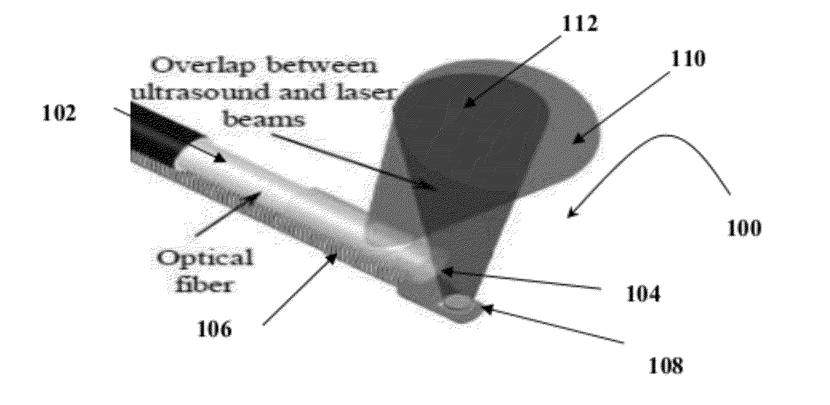
[0014]The optical unit is comprised of one or more optical fibers,
optical fiber bundles or a combination of both and one or more
light delivery systems mounted at the distal end of one or more optical fibers, optical bundles or combination of thereof. In one aspect one or more optical units are used for imaging purposes delivering the short pulses of radiation at desired spectral range into a lumen and emitting the light near orthogonally to the integrated
catheter's longitudinal axis to generate ultrasound
waves from tissues due to absorption of radiation and consequent
thermal expansion of heated areas of arterial tissues. In one aspect the one or more optical units based on a single optical
fiber, optical bundle or a combination thereof may rotate around a longitudinal axis of the catheter. In another aspect one or more optical units are used for therapeutic purposes delivering a high-power CW radiation or quasi CW radiation at desired spectral range about the distal end of the catheter near orthogonally to the integrated catheter's longitudinal axis to irradiate the
pathology leading to
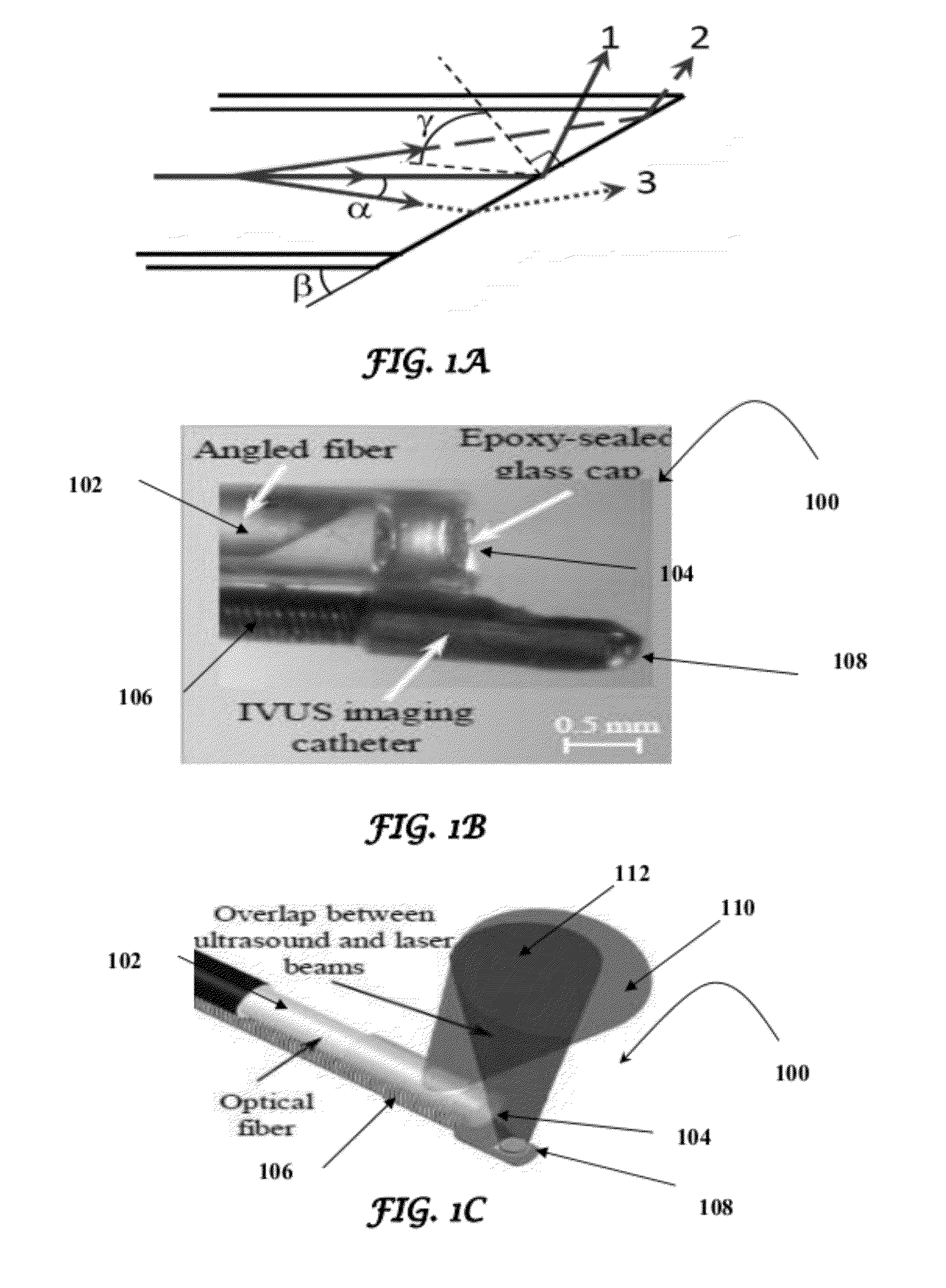
tissue necrosis. A light-transparent tube comprising a sealed distal end and an open proximal end enclosed one or more optical unit's distal ends such that lumen content cannot reach the distal ends of the optical units. In one aspect, related to a side fire fiber-based catheter, the light transparent tube traps a medium such as gas
enclosure to create a difference in the
refractive index between the optical unit's material, and the gas was entrapped around the distal end of the optical fiber that was polished at a certain angle to redirect the light from the polished surface using
total internal reflection effect. In another aspect, related to a micro-optic-based catheter, the distal end of the optical fiber is polished near orthogonally to the integrated catheter's longitudinal axis and emitted light is redirected at the desired angle by one or more optical elements such as micro-mirror, micro-
prism, etc. or in any combinations, and a light-transparent tube traps a medium such as
saline to avoid emitted
radiation attenuation due to interaction with lumen content before light redirection. In both aspects, the light-transparent tube is also used to protect a patient from possible broken-off parts of the catheter. A fixture that is
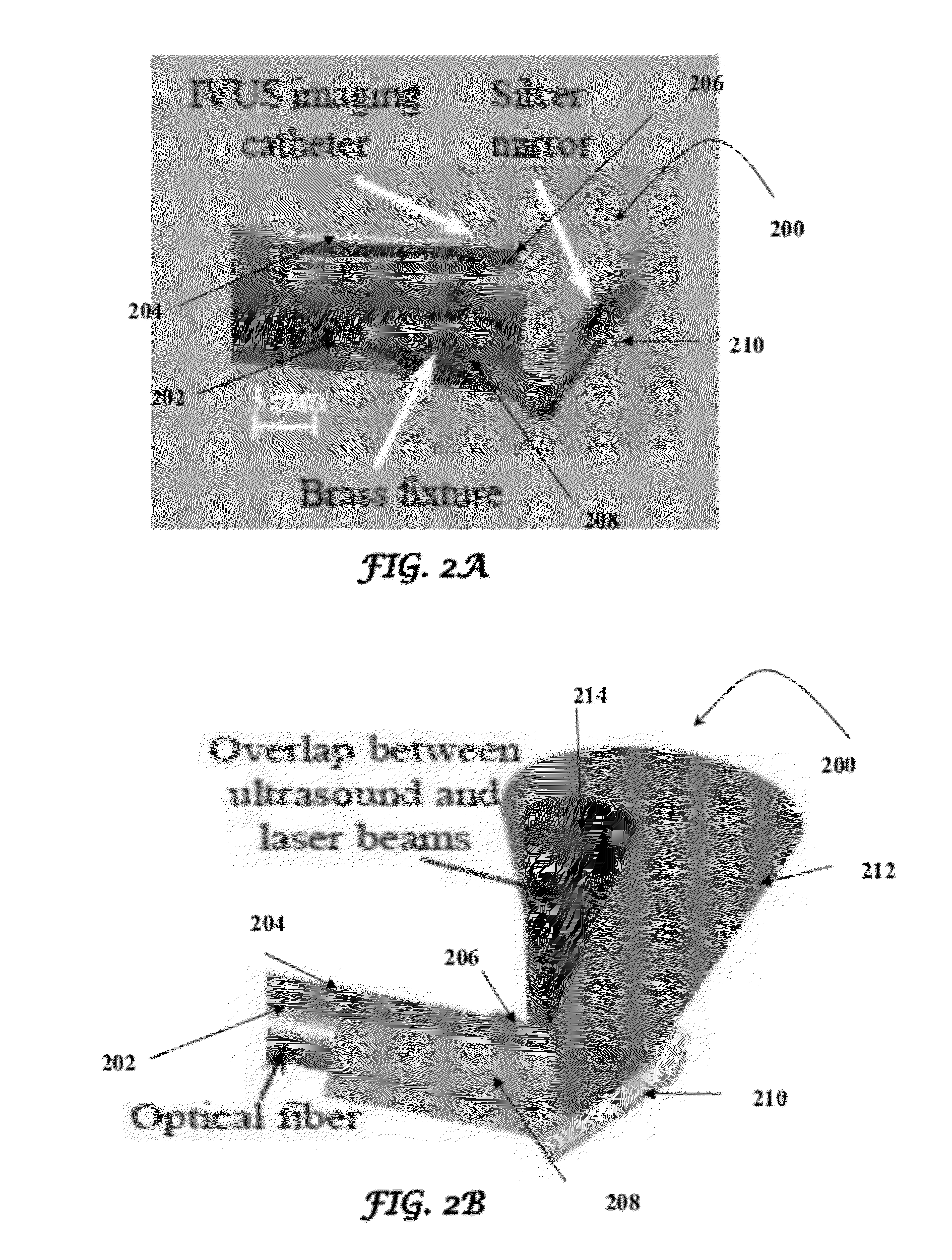
solid near distal end of the device and flexible along the integrated catheter is used to assemble the ultrasound units, optical units or both at the distal end to provide maximum overlap between one or more ultrasound beams emitted by one or ultrasound units and one or more light beams emitted by one or more optical units so the design of the fixture is suitable to concentrate the light in the area where the ultrasound
waves propagate, to encapsulate the parts of the device to make integrated catheter round in cross-section, miniature and safe, and to be used as drive for cross-sectional and longitudinal scan of the vessel lumen.
[0017]In one aspect at least one
ultrasound imaging and therapeutic unit of the present invention is capable of transmitting an
ultrasound wave about the distal end of the catheter and can irradiate an
artery with
pulsed ultrasound waves with consequent detection of the reflected and scattered ultrasound waves in a tissue. The one or more
ultrasound imaging and therapeutic units can provide pulses of ultrasound waves with duration in a range of 1
nanosecond through 1
microsecond with a consequent detection of the ultrasound waves reflected and / or scattered from the tissues. In a specific aspect a central frequency of one or more
ultrasound imaging and therapeutic units is chosen to provide a required resolution and a
penetration depth to image the
artery and nearby tissues and plaques. In another aspect the one or more ultrasound imaging and therapeutic units can irradiate the
artery with a
long pulse or CW
ultrasound wave to provide a
therapeutic effect. The central frequency of one or more ultrasound imaging and therapeutic units is chosen to provide an
ultrasound wave capable of performing a therapy. The present invention allows for varying the duration of the pulses and a
duty cycle of ultrasound waves as required for acoustic therapy.
[0019]In one aspect a
light delivery system is based on micro-
optics. The micro-
optics is attached to the distal end of the one or more optical units. An optically transparent tube sealed on the distal end is mounted on the distal end of the one or more optical units as a separation between the micro-optics and imaged tissue. The tube is filled by a medium such as
saline or water to reduce the
radiation loss during
light transmission. In another aspect a light
delivery system utilizes the total internal effect. The distal end of optical fibers is polished at a certain angle to redirect light to almost near-right angle relative with respect of the longitudinal axis of the catheter. The optically transparent tube sealed on the distal end is mounted on one or more optical units hermetically to trap a medium such as gas near the distal end of optical units to create a difference in the
refractive index between the optical unit's material and the entrapped medium. In both aspects the optically transparent tubes in both designs of the present invention is also mounted on the distal end of the one or more optical units to prevent mechanical damage of the artery. In one aspect the one or more optical units emits short pulsed light with a high
fluence to perform a photoacoustic imaging. In another aspect the one or more optical units are capable of transmitting the CW or the long-
pulse radiation to perform a
light therapy.
[0020]In one aspect the
pulsed laser is coupled with the proximal end of one or more optical units to irradiate the
target tissue at one or more wavelengths, wherein the wavelengths of
electromagnetic radiation are chosen to provide the best
optical contrast. In another aspect of the device of the present invention the
CW laser is coupled with proximal end of the one or more optical units to irradiate target tissues at one or more wavelengths. Both pulsed and
CW laser sources can be coupled with same or different optical units as it is required for necessary procedure. In yet another aspect of the device of the present invention the imager is capable of providing the reconstructed distributions of ultrasound impedances, optical absorption and shear
elastic modulus and of instructing a user to perform an acoustic and / or an optical therapy.


