Amatoxin-Armed Tartget-Binding Moieties for the Treatment of Cancer
- Summary
- Abstract
- Description
- Claims
- Application Information
AI Technical Summary
Benefits of technology
Problems solved by technology
Method used
Image
Examples
example 1
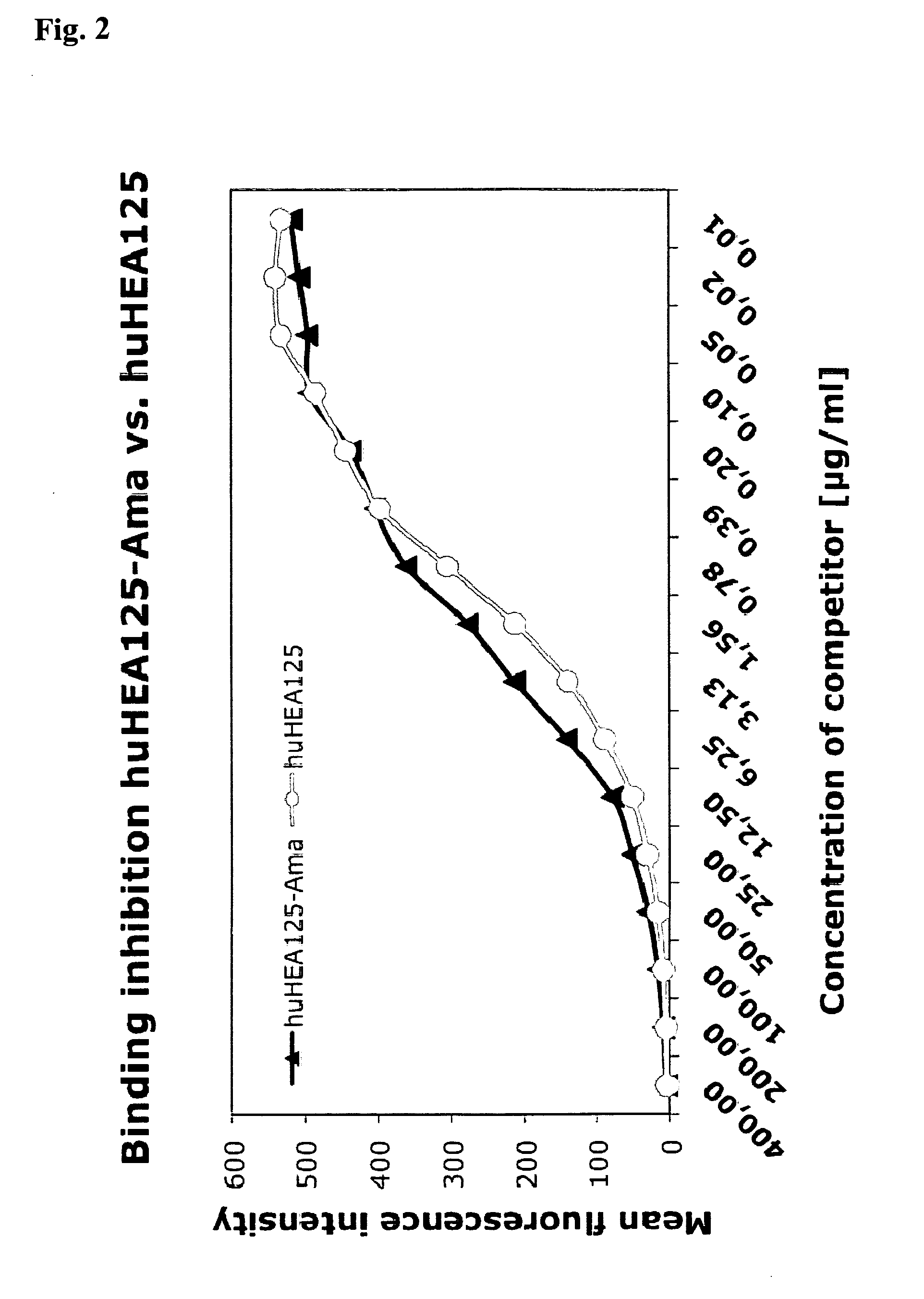
Comparison of Binding Affinities to Target Cells Between Antibody huHEA125 and Antibody Toxin Conjugate amanitin-huHEA125
[0099]1.1 Chimeric Antibody huHEA125
[0100]Several years ago, the inventors have established a hybridoma cell line secreting the anti-EpCAM mouse monoclonal antibody HEA125 (Moldenhauer et al., 1987; Momburg et al., 1987). Using molecular biology techniques this hybridoma line was reconstructed to produce a chimeric version of the antibody consisting of the mouse variable domains hooked up to human kappa constant light chain and human IgG1 constant heavy chain. The resulting antibody huHEA125 binds to EpCAM-expressing cells with high affinity (Kd=2.2×10⁻⁹ M) and high specificity. The gene sequence and the amino acid sequence of huHEA125 immunoglobulin are shown below:
[0101]huHEA125 Heavy Chain
Peptide sequence heavy chain, membrane bound form (IGHV / IGHD / IGHJ / IGHG1; IGHG1 is underlined) (SEQ ID NO: 1):
EVKLLESGGGLVQPGGSLKLSCAASGFDFSRFWMTWVRQAPGKGLEWIGEINLDSSTINYTPSLKD...
example 2
Surface Expression of EpCAM Antigen on Various Carcinoma Cell Lines Detected by Indirect Immunofluorescence
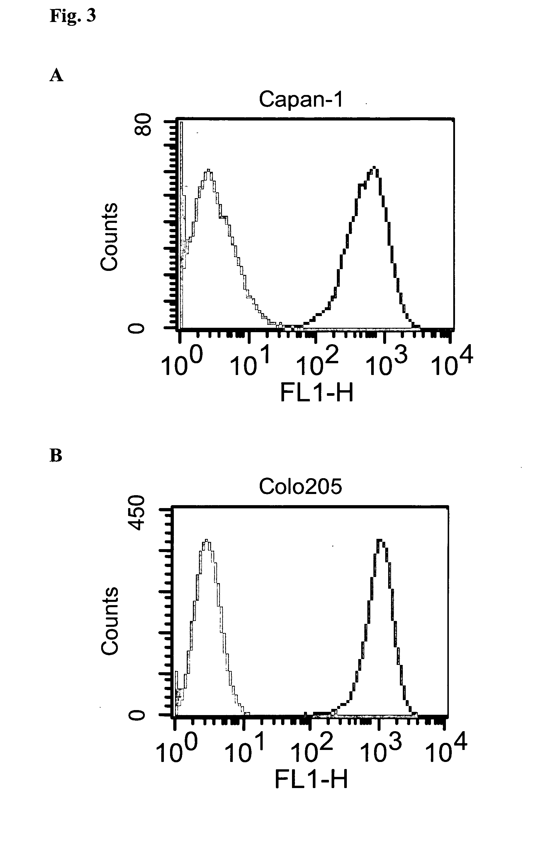
[0112]Cell lines Capan-1, Colo205, OZ, MCF-7, BxPC-3 and PC-3 were first incubated with either huHEA125 or Xolair®. After washing, binding of the primary antibody was visualized by FITC-labelled F(ab′)2 goat anti-human IgG (H+L) as second step reagent. The results are shown in FIG. 3A (Capan-1), FIG. 3B (Colo205), FIG. 3C (OZ), FIG. 3D (MCF-7), FIG. 3E (BxPC-3), and FIG. 3F (PC-3). The grey-shaded histograms in the left side of each diagram show the results obtained with control antibody Xolair®; the histograms having a white area in the right side of each diagram show the results obtained with antibody huHEA125.
example 3
Induction of Carcinoma Cell Proliferation Inhibition by Amanitin and Amanitin / Antibody Conjugates
3.1 Carcinoma Cell Lines
[0113]The following carcinoma cell lines were used for growth inhibition studies:
Capan-1, BxPC-3human pancreatic adenocarcinomaMCF-7human breast adenocarcinomaColo205human colon cancer metastasisOZhuman cholangiocarcinomaPC-3human prostate adenocarcinoma
3.2 Proliferation Inhibition Assay
[0114]Inhibition of cell growth by amanitin-IgG conjugates was determined by incorporation of [3H]-thymidine. Serial dilutions of amanitin-huHEA125, amanitin-Xolair and free amanitin in complete medium (RPMI 1640 supplemented with 10% heat-inactivated FCS, 2 mM L-glutamine and 1 mM sodium pyruvate) ranging from 2×10⁻⁵ M to 6×10⁻¹³ were prepared in triplicates in a volume of 100 μl in the wells of a 96 well flat-bottom tissue culture microtiter plate. Cells were added in a volume of 50 μl per well at a density of 2×10⁴ per ml. Plates were incubated in a humidified atmosphere at 37° ...
PUM
 Abstract
Description
Claims
Application Information
 - R&D
- Intellectual Property
- Life Sciences
- Materials
- Tech Scout
- Unparalleled Data Quality
- Higher Quality Content
- 60% Fewer Hallucinations
Browse by: Latest US Patents, China's latest patents, Technical Efficacy Thesaurus, Application Domain, Technology Topic, Popular Technical Reports.
© 2025 PatSnap. All rights reserved.Legal|Privacy policy|Modern Slavery Act Transparency Statement|Sitemap|About US| Contact US: help@patsnap.com


