
Systems and Methods for Object Identification

a technology of object identification and object identification, applied in image analysis, image enhancement, instruments, etc., can solve problems such as artifacts that cannot be considered clinically significant, and achieve the effect of minimizing the impact of color variation

Inactive Publication Date: 2012-08-16
CHARLES RIVER LAB INC
View PDF7 Cites 17 Cited by
  • Summary
  • Abstract
  • Description
  • Claims
  • Application Information

AI Technical Summary

Benefits of technology

"The present invention provides a system and method for identifying objects in images of biological samples. The system can segment tissue regions and objects in images of samples with significant color differences, while ignoring inconsequential color differences. The system can detect and segment objects using a learned model or user-defined objects, and can handle color variation and lab-to-lab, specimen-to-specimen, and scanner-to-scanner variation. The system can create data structures with the detected objects at approximately the same location in the image domain and can select at least some of the detected objects for further analysis. The technical effects of the invention include improved accuracy and robustness in identifying objects in biological samples and improved automated analysis of images."

Problems solved by technology

When the diagnostician is reviewing a set of hundreds or even thousands of samples, human fallibility may cause artifacts to be deemed clinically significant features and vice versa.

Method used


Image

  • Systems and Methods for Object Identification
  • Systems and Methods for Object Identification
  • Systems and Methods for Object Identification

Examples


Embodiment Construction

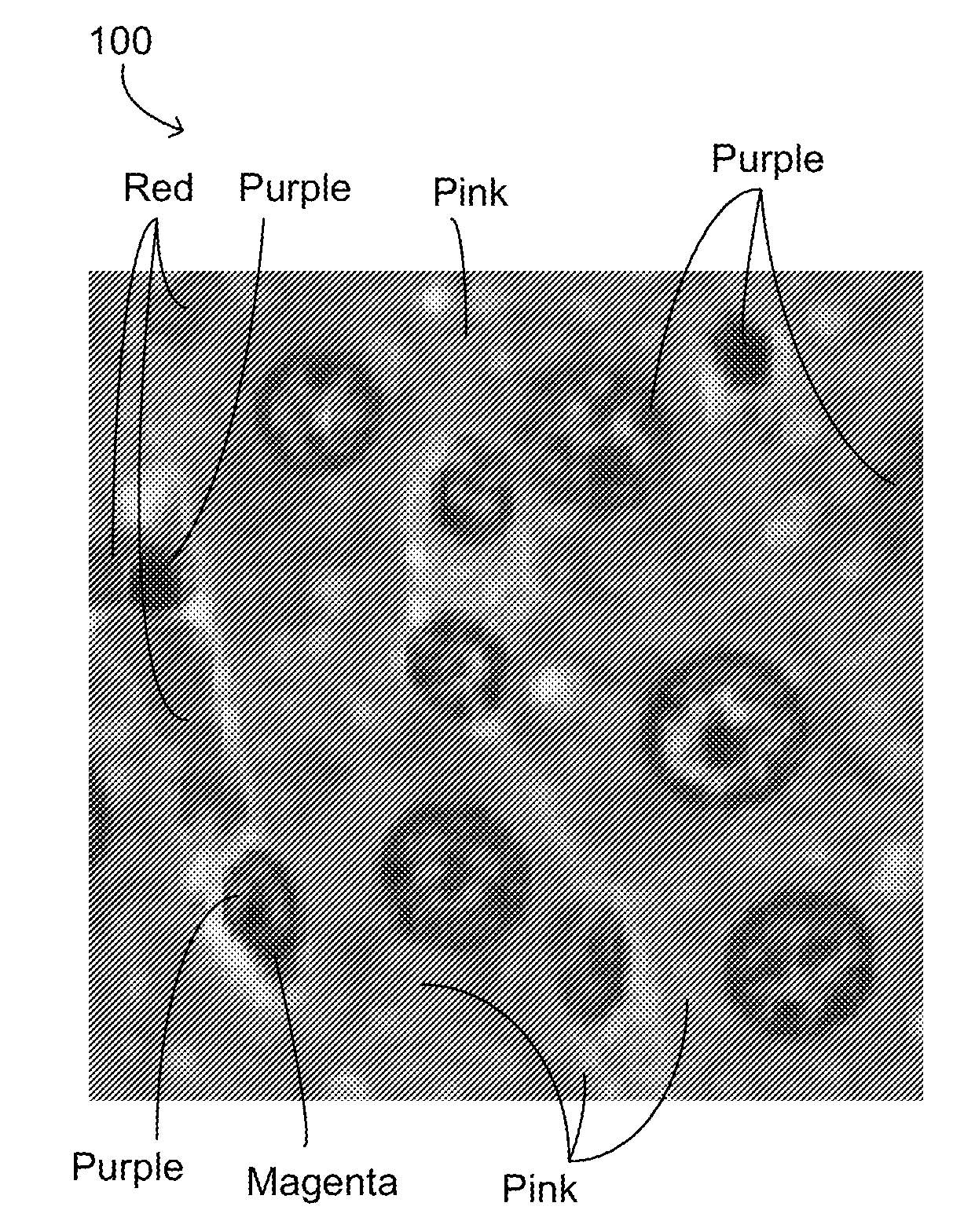
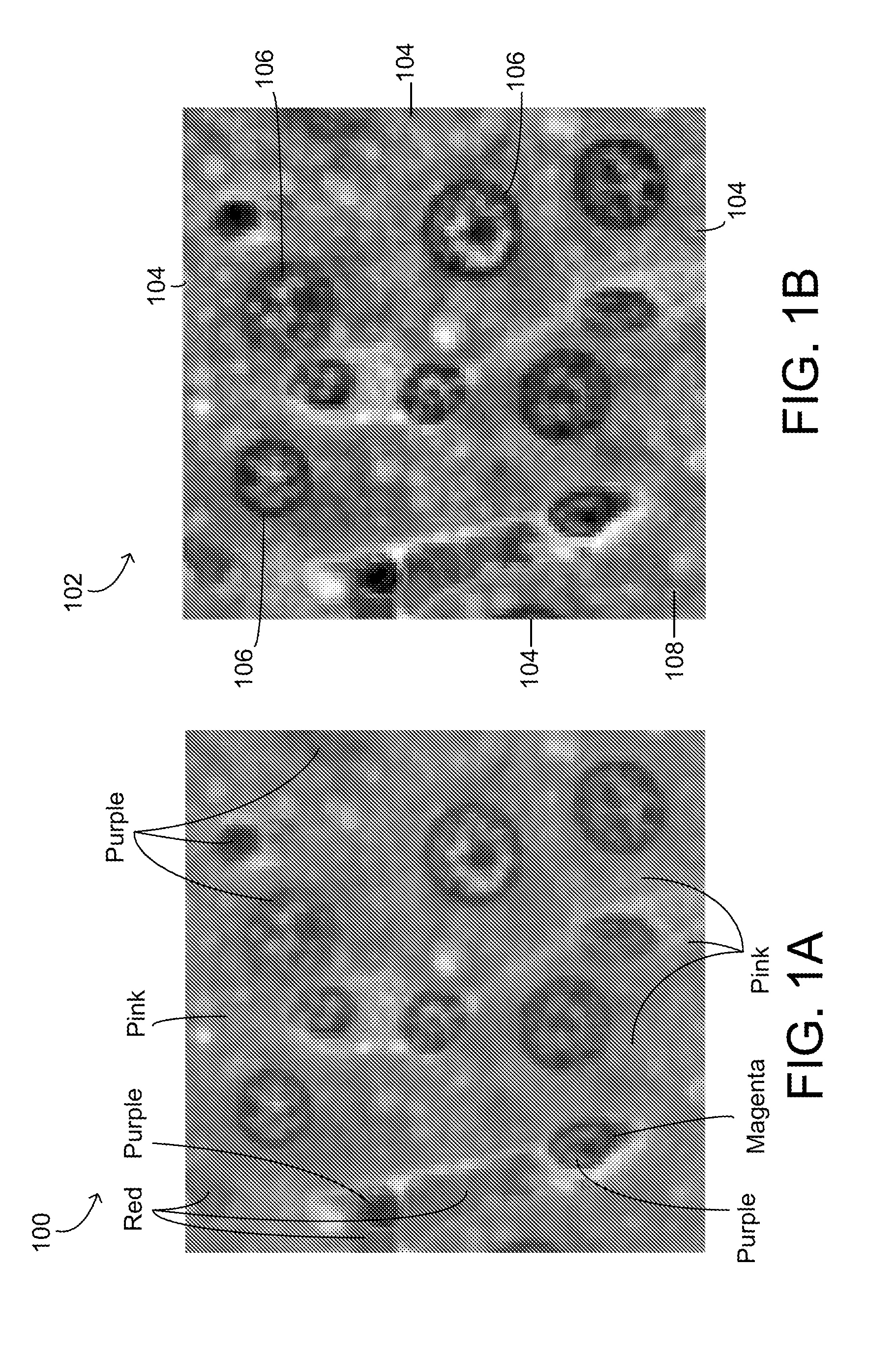
[0021]Embodiments of the present invention provide a system for identifying objects in images. The input images may be multi-channel or grayscale images from any of a number of sources. One common source is images of stained microscope slides. The slides may be stained according to a number of protocols, such as the hematoxylin and eosin (H & E) and the immunohistochemistry (IHC) protocols. The images may be defined in any of a number of color spaces, including, but not limited to, RGB, L*a*b, and HSV. The following discussion assumes an RGB image of a stained microscope slide, but it is to be understood that this example does not limit the domain of applicability of the current invention in any manner.

[0022]A toxicologic pathology study involves the administration of a drug to a plurality of animals, usually in various dose groups, including a control group. After the animals are sacrificed, typically one or more tissues are sectioned, stained, and mounted on microscope slides. The...


PUM


Abstract

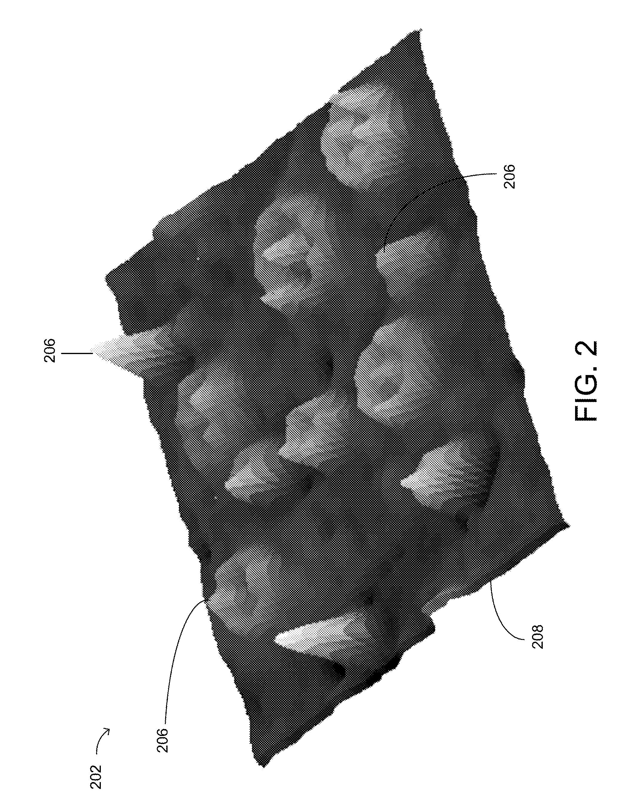
Systems and methods for object identification. Objects in a color image of a biological sample are identified by using a signal function to transform the color image into a single-channel image with localized extrema. The localized extrema may be segmented into objects by an iterative thresholding process and a merit function may be used to determine the quality of a given result.

Description

FIELD OF THE INVENTION[0001]The present invention relates to systems and methods for identifying objects in an image, and in particular multi-channel images.BACKGROUND OF THE INVENTION[0002]Toxicologic pathology is the study of functional and structural changes induced in cells, tissues and organs by external stimuli such as drugs and toxins. Toxicologic studies are helpful to assessing the safety of drugs, vaccines, and other chemicals. A typical toxicologic study involves the controlled administration of at least one substance to a population of test animals. Tissue is harvested from the population using surgical processes such as necropsy. The harvested tissue is typically stained to improve the visibility of various tissue components. After processing, the tissue is mounted on a transparent substrate for viewing or digital imaging. By viewing the specimens, a diagnostician can identify the effects of the administered substance on the members of the test population.[0003]The diag...

Claims


Application Information

Patent Timeline
Patent Type & Authority Applications(United States)
IPC IPC(8): G06K9/62G06K9/46G06K9/34G06K9/00
CPCG06K9/0014G06V20/695G06F18/24G06T7/0012G06T2207/30004
Inventor MEHANIAN, COUROSHLORENZEN, PETER J.LEE, MATTHEW T.ZHU, YANNING
Owner CHARLES RIVER LAB INC
Learn More