
Apparatus and method for affixing frozen tissue sections to glass or membrane microscope slides

a microscope slide and tissue technology, applied in the field of molecular biology and microbiology, can solve the problems of tissue section improper folding onto the slide, many problems, and the learning curve of the method is too high to achieve the effect of properly performing the method without tissue section folding, reducing the need for reagents, and increasing economic efficiency in the laboratory

Inactive Publication Date: 2012-03-08
LEININGER ERIC JEFFORDS +1
View PDF5 Cites 7 Cited by
  • Summary
  • Abstract
  • Description
  • Claims
  • Application Information

AI Technical Summary

Benefits of technology

[0014]The invented apparatus allows tissue sections to remain flat and adherent on the glass or membrane slide with little difficulty. Further, the embedding medium is removed prior to affixing the tissue section to the slide so that micro-folding of tissues within a section does not occur and coagulation of embedding medium does not need to removed prior to laser micro dissection, enabling greater RNA stability. In addition, thick tissue sections (>25-200 μM) easily adhere to the slide. Also, many sections can placed on one slide decreasing necessary reagents and increasing economic efficiency in the laboratory.

Problems solved by technology

Currently, the “swooping heat method” is the most widely used, and while it is inexpensive relative to the “UV method”, there is a steep learning curve in order to perform this method properly without tissue section folding.
If the tissue sections are curled, which is dependent on the temperature gradient between the cryostat and the room, or dependent on the incident angle between the slide and ribbon of sections, the tissues will improperly fold onto the slide.
This can cause many problems.
For example, if the tissue sections are folded, the architecture of the tissue, upon subsequent processing and imaging, is difficult to interpret.
In addition, improper tissue folding will tend to have the sections fall off the slide as the less tissue surface area means less electrostatic interaction between the tissue and the slide upon subsequent processing.
Finally, if the angle of the slide used to swoop down and pick up the tissue ribbon is grossly inaccurate or the sections are extensively curled then (for example, when a beginner is attempting to perform the procedure) then the tissue sections can become stacked one on top of the other and further processing would be ill-advised.
Overall, in order to perform the “swooping heat method” without improper tissue folding, it takes tens-hundreds of hours of training.
Besides improper tissue folding, another problem with the “swooping heat method” is that, if done improperly, large amounts of tissue sections can be lost.
This is because the both the ribbon of tissue sections and glass slide are lengthwise in nature and it is not economically feasible to collect only one section per slide for most experiments.
Therefore, if the swooping process does not accurately lay down the sections in the desired manner, swaths of tissue sections are lost, and the user can lose valuable data.
Further, though the tissue sections may be small in surface area relative to the slide and multiple samples are being collected on one slide, the procedure still uses many slides as the number of tissue sections that can normally fit on one slide in one horizontal plane is limited.
The more slides that are used the more reagents (for example antibodies) are necessary in order to process the tissue further down the line, raising the expense necessary to perform the procedure.
In addition to improper tissue folding and lost sections, the “swooping heat method” may induce micro-folding of tissue structures within an individual section.
Another problem with the “swooping heat method” is that sections >25 μM (for performing whole-mount immunohistochemistry, or for collecting larger tissue volumes for laser micro dissection) are less able to maintain their adhesion, due to the large amount of embedding medium that interrupts the electrostatic interaction of the tissue and the slide.
While the problems inherent in the swooping “heat method” for glass slides are the same for membrane slides, there is an additional complication that arises for membrane slides.
This complication arises because the subsequent processing of the tissue sections for laser micro dissection involves the goal of RNA stabilization.
However, because the water soluble embedding medium, present on the membrane with the tissue section, needs to be removed so that it does not inhibit laser penetration of the tissue, the subsequent processing of the tissue must involve addition of an aqueous solution.
However, the “UV method” has some problems as well.
For example, very few sections can fit on one slide making the collection of tissue slower than the “swooping heat method”.
Further , the equipment needed is quite specific and expensive including special tape that can operate at 0° to −40° C., special slides coated with an adhesive necessary to transfer the section, and a low temperature, cryostat-embedded UV light used for cross linking Additionally, these packaged slides are not Rnase free.

Method used


Image

  • Apparatus and method for affixing frozen tissue sections to glass or membrane microscope slides
  • Apparatus and method for affixing frozen tissue sections to glass or membrane microscope slides
  • Apparatus and method for affixing frozen tissue sections to glass or membrane microscope slides

Examples


Embodiment Construction

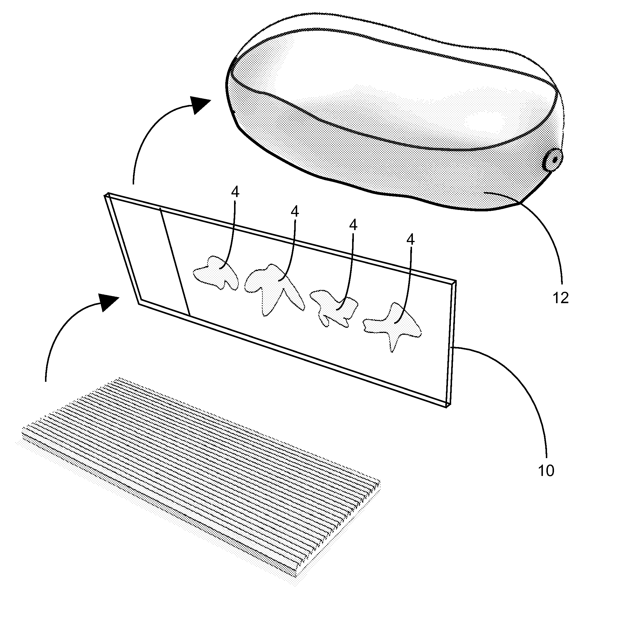
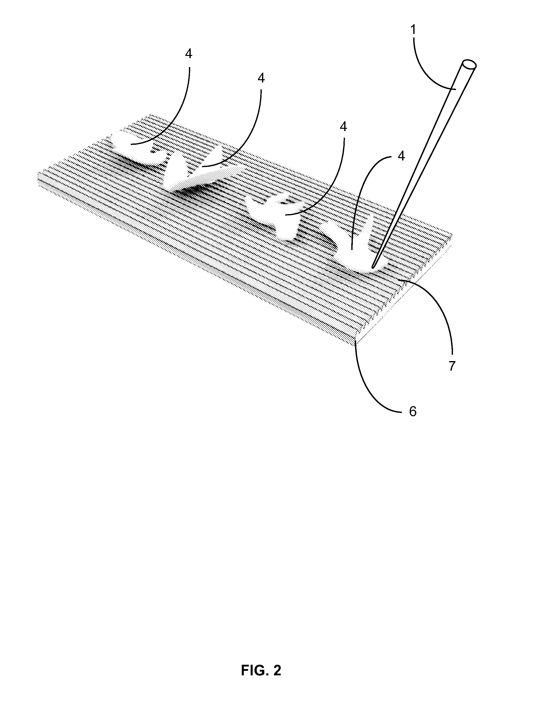
[0023]The function of this apparatus is to affix frozen tissue sections to glass or membrane microscope slides. The apparatus contains several parts; teasing implement(s) for removing the tissue section from the surrounding embedding medium, an affixing block with one or more affixing surfaces which enables the sections to be flattened and transferred to the slide and a heating element which induces transference of the section from the affixing block to the slide.

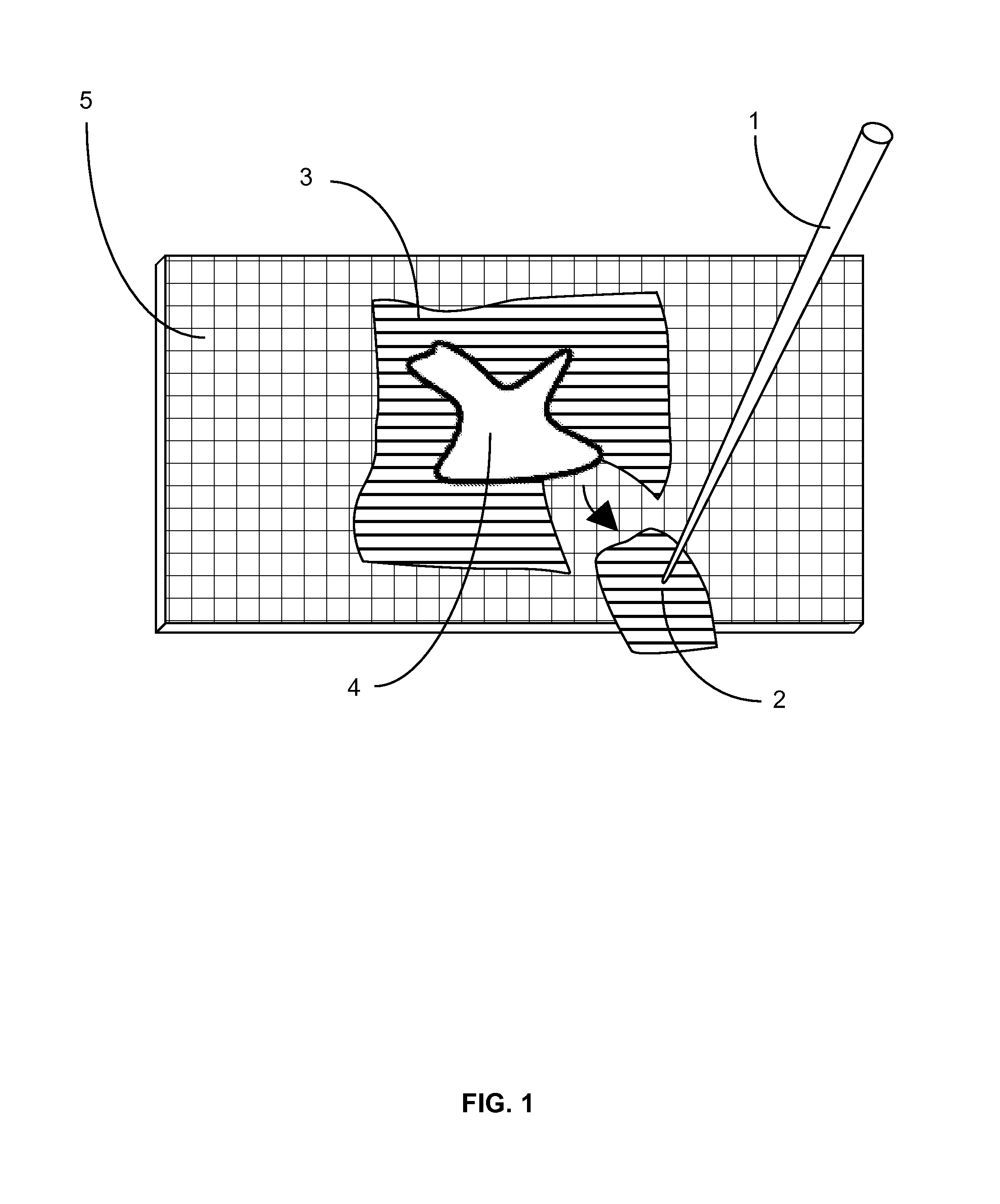
[0024]First, after cutting each tissue section, an individual first removes the surrounding embedding medium from the tissue section (FIG. 1). This is done inside the cryostat and with temperature-equilibrated teasing implement(s). The teasing implement (FIG. 1; 1) is small enough to be easily manipulated by hand and has a pointed tip (FIG. 1; 2) so that the surrounding embedding medium (FIG. 1; 3) can be removed from the tissue section (FIG. 1; 4) on a nominal surface (FIG. 1; 5), which provides support, inside the cryosta...


PUM


Abstract

This invention is an apparatus and method for affixing frozen tissue sections to microscope slides. It has three elements; teasing implements, an affixing block and a heating element. The elements are used inside a cryostat prior to processing for molecular biology procedures such as immunohistochemistry or laser micro dissection. The tissue sections are collected onto an affixing block which has indentations that decrease the contact surface area while providing structural support. The tissue sections are then transferred onto a slide via transient application of a heating element to the back of slide. The invention decreases folding of tissue sections, increases morphological consistency, enables the placement of many sections on one slide and enables the adherence of tissue sections with greater thickness.

Description

FIELD OF INVENTION[0001]The suggested field of invention for this invention is CHEMISTRY: MOLECULAR BIOLOGY AND MICROBIOLOGY CLASS #435 and the suggested subclasses are APPARATUS #283.1 and subclass IMMUNOHISTOCHEMICAL ASSAY #935.BACKGROUND ART[0002]In order to perform some forms of immunohistochemistry and laser micro dissection, there is a relatively common technique for collecting the tissue in molecular biology laboratories. First, a region of interest (ROI) is isolated through resection of animal body part. Second, the ROI is placed inside a frozen tissue embedding medium. This embedding medium usually contains polyethylene glycol and is water-soluble. Third, the embedding medium and ROI themselves are placed within a mold. Finally, after placing the mold in contact with liquid nitrogen, the ROI and medium is rapidly frozen. After being frozen, the ROI / embedding medium complex structurally supports the sample for subsequent sectioning at below freezing temperatures.[0003]In ord...

Claims


Application Information

Patent Timeline
IPC IPC(8): G01N33/48C12M1/00
CPCG01N1/06G01N1/36G01N1/286
Inventor LEININGER, ERIC JEFFORDSFERNANDO, WARNAKULASURIYA AKASH
Owner LEININGER ERIC JEFFORDS
Learn More