Insertion of medical devices through non-orthogonal and orthogonal trajectories within the cranium and methods of using

Active Publication Date: 2012-02-23
HUA SHERWIN
View PDF10 Cites 60 Cited by
  • Summary
  • Abstract
  • Description
  • Claims
  • Application Information

AI Technical Summary

Benefits of technology

[0030]The improved method involves modification of implantable devices to specific sizes and shapes so that one or several can be inserted simultaneously through a single entry site in the cranium by altering the insertion angle of each unit. The individual units are inserted orthogonally and / or nonorthogonally relative to the surface of the cranium tangent to the singular common entry site. The individual units may be physically connected through a connector head at the common entry site, thereby sharing electronics, power, and other attributes. Additionally, in some embodiments, the distal tip of the shaft and the shaft of the device may be configured so that the devices are insertable directly. By insertable directly it is meant that no or few other tools or instruments are needed to make the entry site and / or the hole through which the implanted device is inserted. For example, the device may be encapsulated in a helical externally threaded screw housing such that the shaft has a sharp distal tip allowing the whole device to pierce through the skin and screw into bone similar to currently used self-drilling cranial plating screws. The self-inserting characteristic enables electrodes to be inserted almost anywhere very quickly in a minimally invasive screw-in or pop-in procedure.
[0032]The thickness of the cranium is limited to a length of 5 mm to 10 mm. If electrodes are inserted straight down, perpendicular (orthogonal) to the surface of the cranium, their lengths would be limited to a maximum of approximately 1 cm. Electrodes longer than 1 cm that are implanted in the cranium orthogonally would protrude through the skull into the brain. Placement of electrodes into brain substance increases the risk of injury to brain and blood vessels both during the time of placement as well as afterwards given the physiologic pulsation of the brain in relation to the cranium as well as during episodes of head trauma which causes acceleration and deceleration movement of the brain in relation to the cranium. Current methods of cortical stimulation place electrodes either epidurally (outside the dura mater) or subdurally (in between the dura mater and arachnoid or epi-arachnoid). Placement of electrodes in either of these locations provides for low impedance stimulation of the brain while maximizing safety. Current methods of placement of cortical electrodes necessitates drilling of a burr hole or craniotomy, both of which pose risks to the patient and commonly require a stay in the intensive care unit to monitor postoperatively.
[0034]Longer electrodes units also allow more components to be integrated within each implant. Larger size allows flexibility in terms of the complexity of the circuitry, communication components, as well as the inclusion of both recording (receiving) and stimulation (transmitting) capabilities. Additionally, multiple electrode contacts can be placed within a single implant with greater ease, i.e. bipolar, tripolar, tetrapolar stimulation or recording within each electrode unit.

Problems solved by technology

In deep brain stimulation, the electrode passes through the cerebral cortex as well as subcortical brain structures to reach the affected deep brain nuclei and therefore risks injury to the intervening healthy brain tissues as well as blood vessels.
These unnecessary yet unavoidable injuries can potentially result in loss of brain functions, stroke, and intracranial hemorrhage.
Although noninvasive, brain-machine interface using EEG signals is currently limited from the significant dampening of the brainwave's amplitude by the cranium.
These procedures necessitate a minimum of an overnight stay in the hospital and pose risk to injury of the brain due to the invasiveness of the techniques.
Additionally these “open” techniques pose special challenges for securing the electrode as most technologies require a lead to exit the hole in the skull.
Current techniques for cortical stimulation also risk the development of scarring of the cortex as well as hemorrhage.
Scarring distorts the normal brain architecture and may lead to complications such as seizures.
Additionally, the placement of devices on the surface of the brain poses risks of hemorrhage.
Thus in the case of a deceleration injury like that seen in traffic accidents or falls, the imperfect anchoring of the electrode and the mass of the electode may cause the electrodes to detach and injure the brain.
Although muscle and other body parts allow the implantation of bion electrodes, the cranium poses a challenge to the bion because the cranium is roughly 1 cm or less in thickness.
However, these patents are not specially adapted for insertion through the skull with multiple components through a single site by means of introducing some components at non-orthogonal angles.
The patent discloses “[t]he microstimulators, of course, may be planted in or near any part of the body, in the brain, a muscle, nerve, organ or other body area” (See 4:24-26) However, no details are provided on how the microstimulators would be or could be implanted into the brain.
Certainly a hypodermic needle cannot be injected through the skull which suggests these stimulators are not designed for such a purpose.
However, neither patent teaches or suggests incorporating the electrode in a screw housing or other component capable of penetrating the skull or cranium (rather than just the skin) for access to the brain's cortex.
However, there is no disclosure of inserting multiple electrodes through the same entry site.
These designs are significantly more invasive than having just one opening in the cranium and continue to carry the risk of electrodes moving with respect to the brain during head injuries.

Method used


Image

  • Insertion of medical devices through non-orthogonal and orthogonal trajectories within the cranium and methods of using
  • Insertion of medical devices through non-orthogonal and orthogonal trajectories within the cranium and methods of using
  • Insertion of medical devices through non-orthogonal and orthogonal trajectories within the cranium and methods of using

Examples


Embodiment Construction

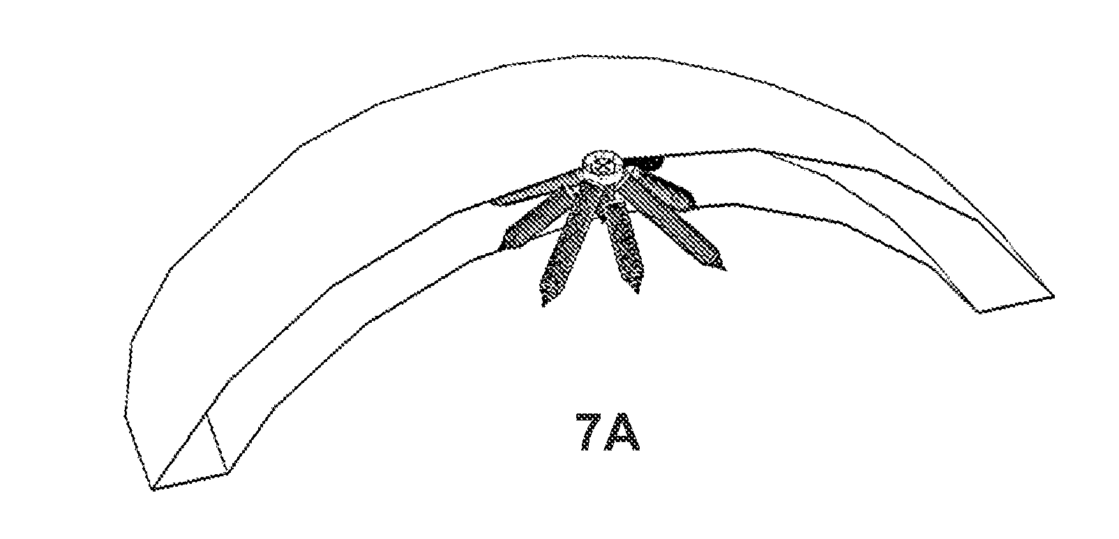
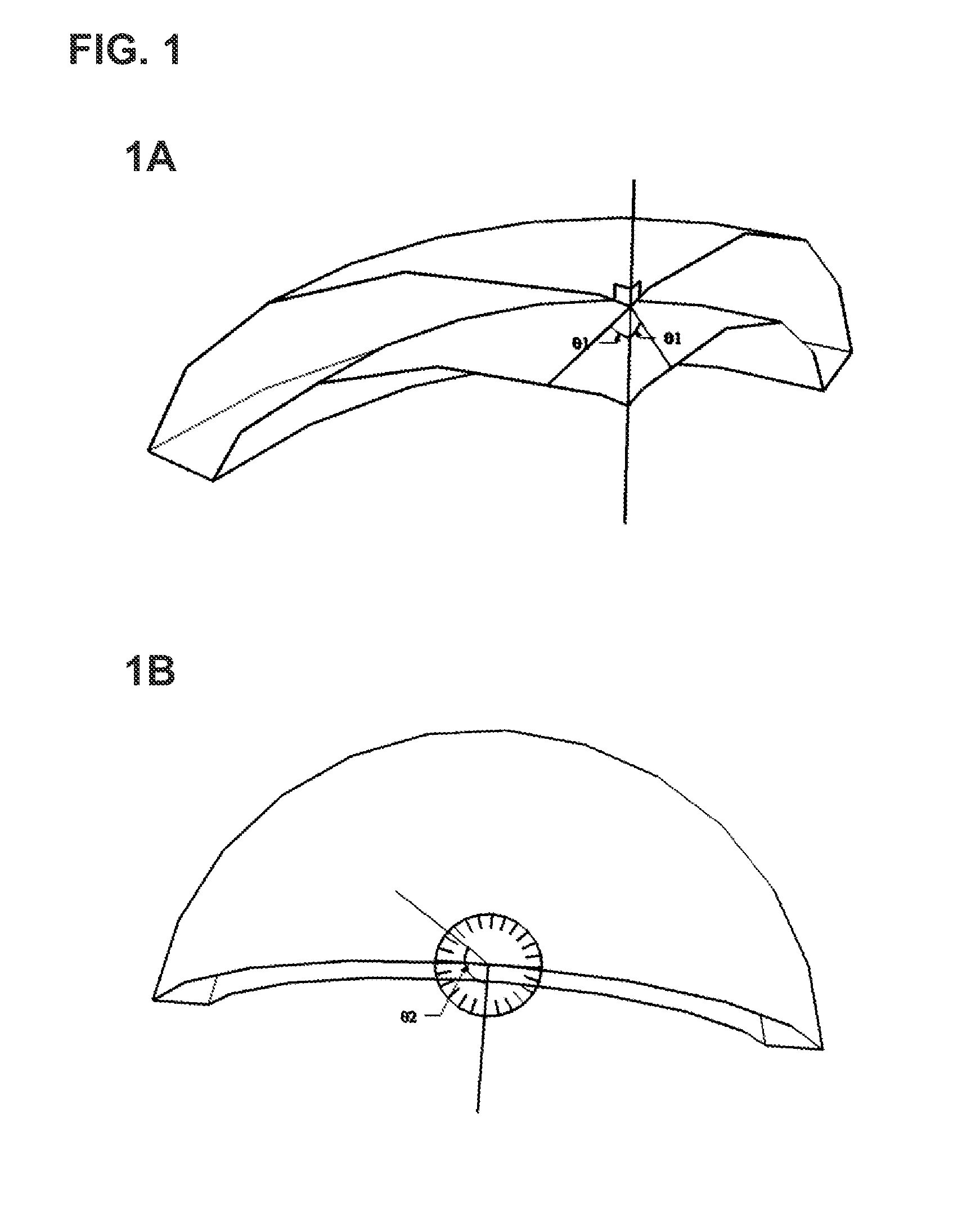
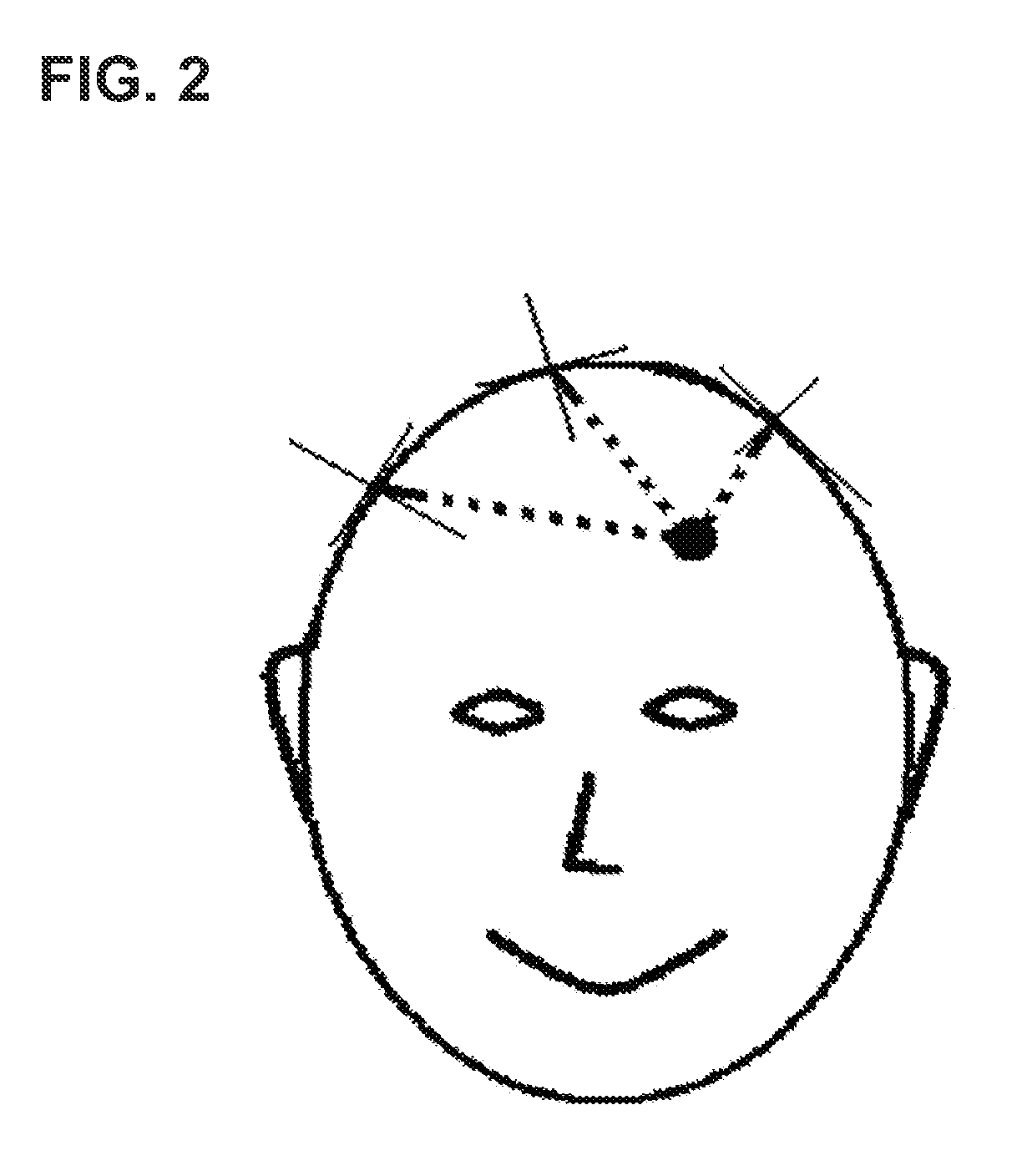
[0053]The present invention and method of its use enables multiple effectors, sensors, and other components to fit through a single entry site to provide improved and / or longer-lasting therapeutic benefits. According to some embodiments this is accomplished by inserting the effectors, sensors, other components, or shafts housing any of these elements at different angles to permit greater subsurface reach given a small surface entry site. As used herein, the term “entry site” includes one or more physically distinct openings, holes, or incisions, within close proximity to one another and taking up a relatively small total area of space consistent with minimally invasive surgical procedures. Thus, an “entry site” may be one opening or hole but is not limited to such. The “entry site” may also be an entry zone, area, or region that encompasses two, three, four, or more distinct openings.

[0054]For each entry site, the stimulator / sensor devices may be inserted at several different axial ...


PUM


Abstract

The invention comprises an elongated device adapted for insertion, including self-insertion, through the body, especially the skull. The device has at least one effector or sensor and is configured to permit implantation of multiple functional components through a single entry site into the skull by directing the components at different angles. The device may be used to provide electrical, magnetic, and other stimulation therapy to a patient's brain. The lengths of the effectors, sensors, and other components may completely traverse skull thickness (at a diagonal angle) to barely protrude through to the brain's cortex. The components may directly contact the brain's cortex, but from there their signals can be directed to targets deeper within the brain. Effector lengths are directly proportional to their battery size and ability to store charge. Therefore, longer angled electrode effectors not limited by skull thickness permit longer-lasting batteries which expand treatment options.

Description

BACKGROUND[0001]1. Field of the Invention[0002]The present invention relates to medical devices, systems and methods for accessing cranial and intracranial structures. Specifically, the invention is directed to altering brain function and treating cranial and intracranial pathology. More specifically, the invention is directed to the surgical implantation of electrodes or other devices within or through the cranium to alter or improve brain function and pathological states such as stroke, seizure, degeneration, and brain tumors. Most specifically, the invention is directed to minimizing surgical methods and risks and maximizing the length of devices that can be implanted within or through the cranium and their ability to hold charge.[0003]2. Description of the Related Art[0004]Electrical stimulation of the brain can improve and ameliorate many neurologic conditions. Examples of the success of brain stimulation include deep brain stimulation for Parkinson's Disease, tremor, dystonia,...

Claims


Application Information

Patent Timeline
IPC IPC(8): A61B5/01A61N1/05A61B6/00A61B8/00A61M5/168A61F7/00A61N5/06A61N5/067A61N1/36A61B19/00A61B5/055A61B6/03A61N2/00A61N7/00A61B5/145A61B5/1459A61B5/03A61B5/00A61B5/375
CPCA61B5/6865A61N1/0531A61N1/0539A61B5/0478A61N1/36064A61N1/36139A61B5/6868A61B5/4836A61B5/4094A61B5/0482A61B5/6882A61B5/4064A61B5/4076A61B5/375A61B5/291A61N1/05A61N1/372A61B17/1695
Inventor HUA, SHERWIN
Owner HUA SHERWIN
Why Eureka
Try Eureka