Imageable rodent model of asthma
a rodent model and asthma technology, applied in animal husbandry, medical preparations, drug compositions, etc., can solve the problems of insufficient th2 response to induce asthma, excessive mucus in the lumen and edema in the submucosa, and epithelial damag
- Summary
- Abstract
- Description
- Claims
- Application Information
AI Technical Summary
Benefits of technology
Problems solved by technology
Method used
Image
Examples
example 1
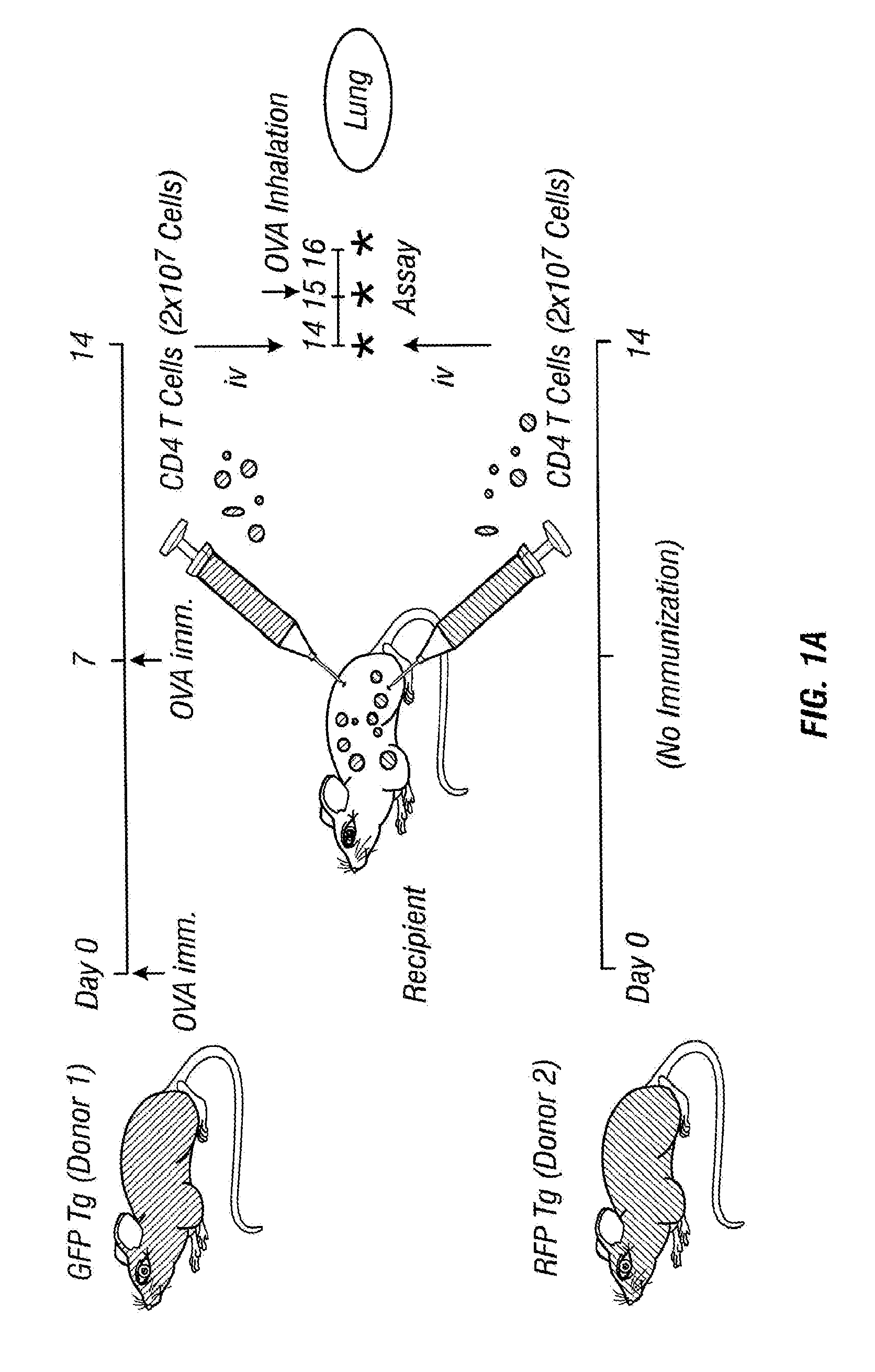
[0049]GFP Tg mice were sensitized with OVA-alum on days 0 and 7.
[0050]Splenic CD4+ T cells from OVA-sensitized GFP Tg and non-sensitized RFP Tg mice were purified and injected into normal C57BL / 6 mice on day 14. The recipient mice were exposed to aerosolized OVA allergen challenge by airway administration on day 15. On day 16, GFP+ and RFP+ CD4+ T cells on the surface of the lung were monitored by OV100 microscopy (FIG. 1a).
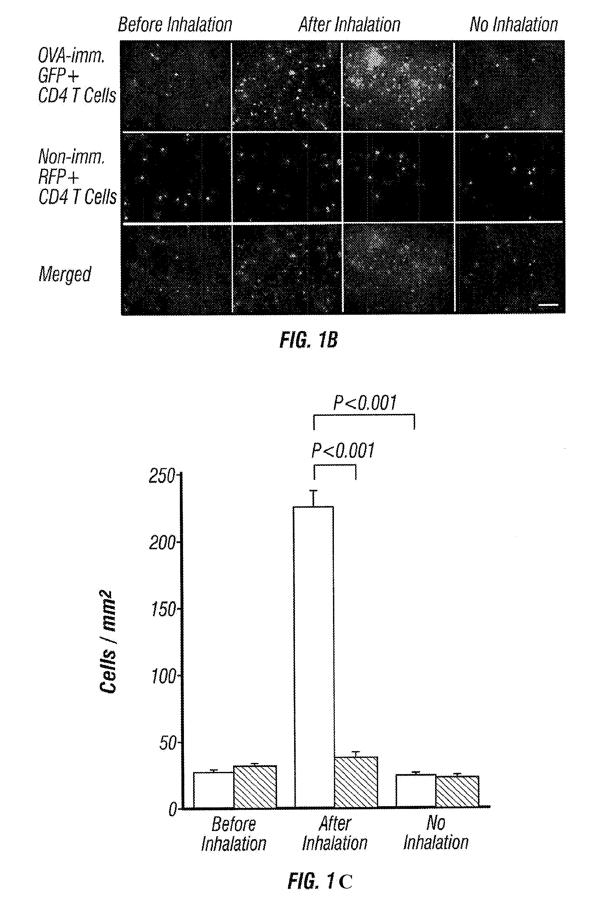
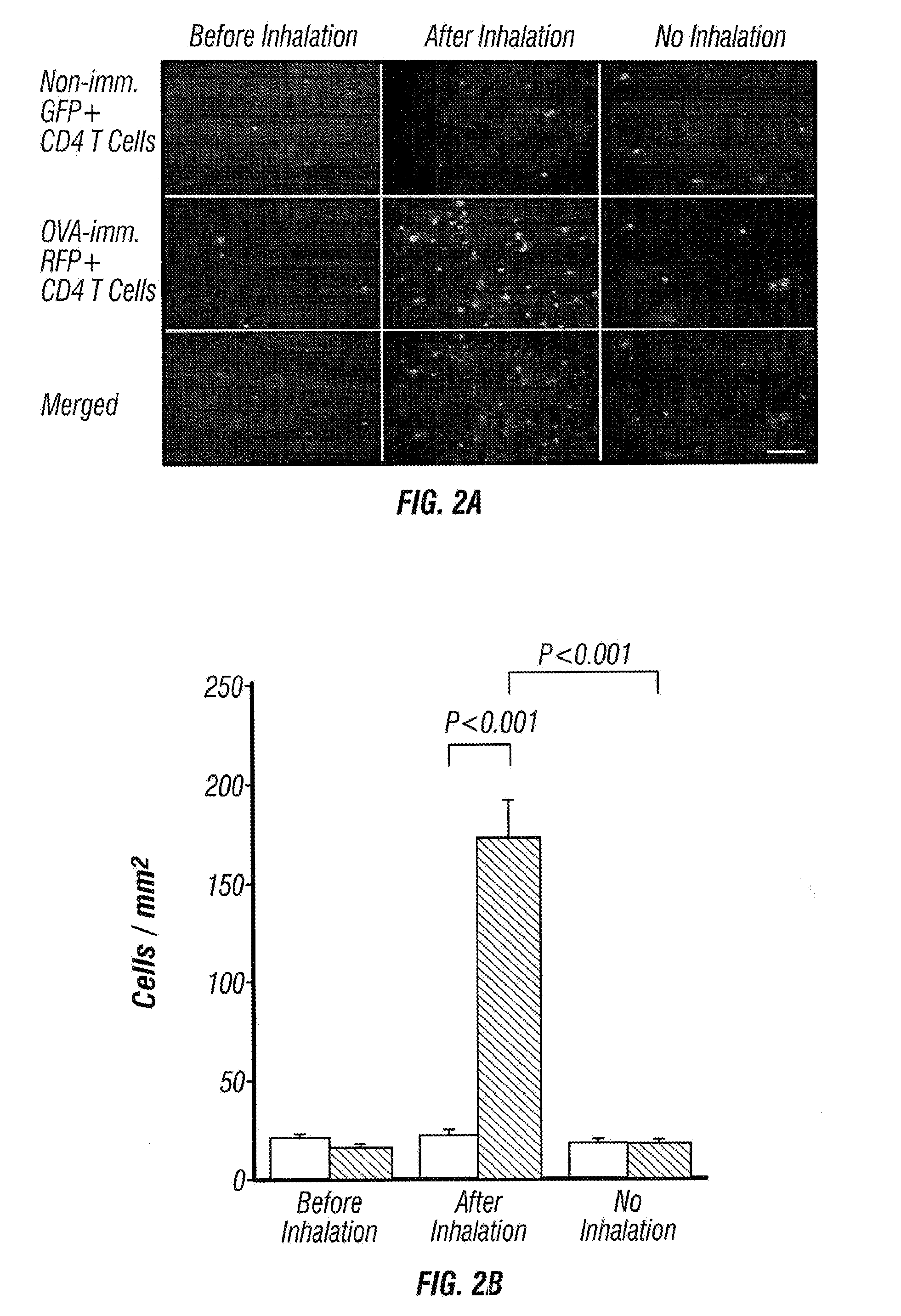
[0051]Immediately after injection, large numbers of transferred cells were accumulated in the lung capillaries (FIG. 1b). Similar numbers of GFP+ and RFP+ cells were detected. One day after cell transfer, there was no significant number of GFP+ and RFP+ cells remaining in the lung. Twenty four hours after OVA inhalation, however, the number of GFP+ CD4+ T cells from OVA-sensitized mice increased significantly and some of them formed foci that look like clusters of CD4+ T cells. On the other hand, the number of RFP+ CD4+ T cells from non-sensitized mice did not in...
example 2
[0054]The time course of CD4+ T cell accumulation in the lung after OVA inhalation was examined.
[0055]Splenic CD4+ T cells from OVA-sensitized GFP Tg mice were injected into recipient C57BL / 6 mice, and the recipient mice were exposed to an allergen challenge as described in Example 1. GFP+ CD4+ T cells on the surface of the lung were monitored at 24 hours (FIGS. 4a and 5a) and 72 hours (FIGS. 4b and 5b) after OVA inhalation by OV100 microscopy. Migration of GFP+ CD4+ T cells into the lung was first detected at 12 hours after OVA inhalation, and the maximum number of CD4+ T cells was detected at 18 to 36 hours after OVA inhalation. While eosinophil infiltration is characteristic in allergic airway inflammation, these results indicate that CD4+ T cell accumulation in the lung after the allergen challenge occurs prior to infiltration of eosinophils, and sustains to at least 72 hours after the allergen challenge.
[0056]This imageable model proves useful to monitor the migration of inflam...
example 3
[0060]To investigate the dynamics of antigen-specific Th2 cells in the asthmatic lung, OVA-specific Th2 cells were induced in vitro from naive CD4+ T cells from GFP Tg×OT2 Tg mice.
[0061]First, we confirmed the accumulation of GFP+ OVA-specific Th2 cells in the lung after an allergen challenge. GFP+ OT2-Th2 cells accumulated in the lung after OVA inhalation more efficiently than CD4+ T cells from OVA-primed mice (FIG. 6). The number of foci with OT2-Th2 cells was much more than that with OVA-primed CD4+ T cells (data not shown). These results indicate that OT2-Th2 cells induced in vitro accumulate in the lung more efficiently after antigen inhalation.
[0062]Next, we performed time course analysis of OT2-Th2 cell accumulation after OVA inhalation. OT2-Th2 cell formed small foci 6 hours after OVA inhalation. The number and size of the foci increased 12 hours after OVA inhalation. GFP+ cell number in non-focus area also increased but not significantly until 12 hours after OVA inhalation....
PUM
 Abstract
Description
Claims
Application Information
 - R&D
- Intellectual Property
- Life Sciences
- Materials
- Tech Scout
- Unparalleled Data Quality
- Higher Quality Content
- 60% Fewer Hallucinations
Browse by: Latest US Patents, China's latest patents, Technical Efficacy Thesaurus, Application Domain, Technology Topic, Popular Technical Reports.
© 2025 PatSnap. All rights reserved.Legal|Privacy policy|Modern Slavery Act Transparency Statement|Sitemap|About US| Contact US: help@patsnap.com


