Microfluidic imaging cytometry
a microfluidic and cytometry technology, applied in the field of microfluidic systems, can solve the problems that egfr mutations cannot account for the responsiveness of egfr kinase inhibitors, and no discovery platform currently availabl
- Summary
- Abstract
- Description
- Claims
- Application Information
AI Technical Summary
Benefits of technology
Problems solved by technology
Method used
Image


Examples
example a
Assay Example
[0099]1) Sample preparation, including cell loading, cell culture in an incubator and media exchange for cell maintenance.
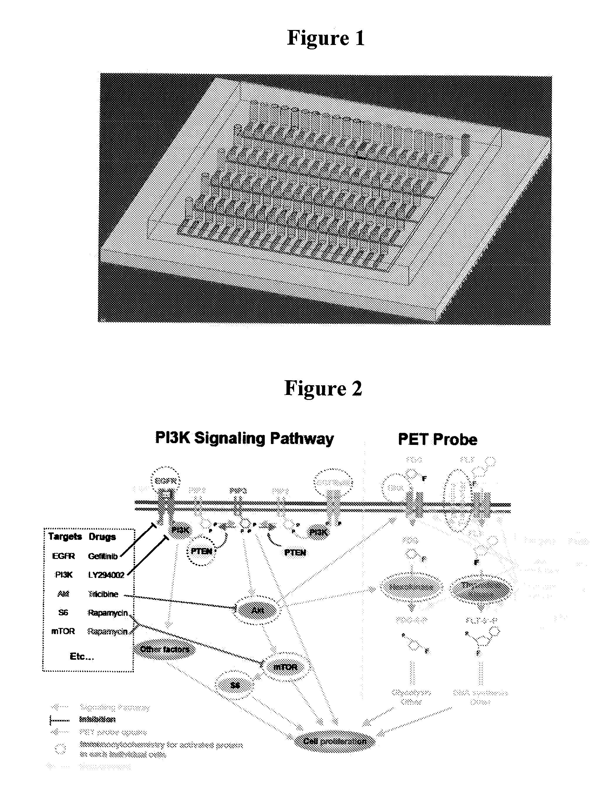
[0100]2) Immunocytochemistry, including cell fixation, permeabilization, and immunostaining.[0101]Multiple signaling nodes including EGFR, EGFvIII, PTEN, pAkt, pmTOR, pS6, and the proliferation marker Ki67, involved in PI3K-Akt-mTOR signaling network can be measured in glioblastoma system.[0102]Multiple signaling nodes including EGFR, ErbB2, PTEN, pAkt, pmTOR, pS6, and the proliferation marker Ki67, involved in PI3K-Akt-mTOR signaling network can be measured in breast cancer system.
Growth Curve of the Living Cell can be Monitored as the Follows:
[0103]The cell culture / assay chip shown in FIG. 8 composed of two types of microchannels responsible for (i) performing 72 cell cultures and assays in parallel and (ii) moisturizing the adjacent cell culture chambers (preventing media evaporation). Meanwhile, we have devoted a significant amount of efforts to...
example b
Data Collection
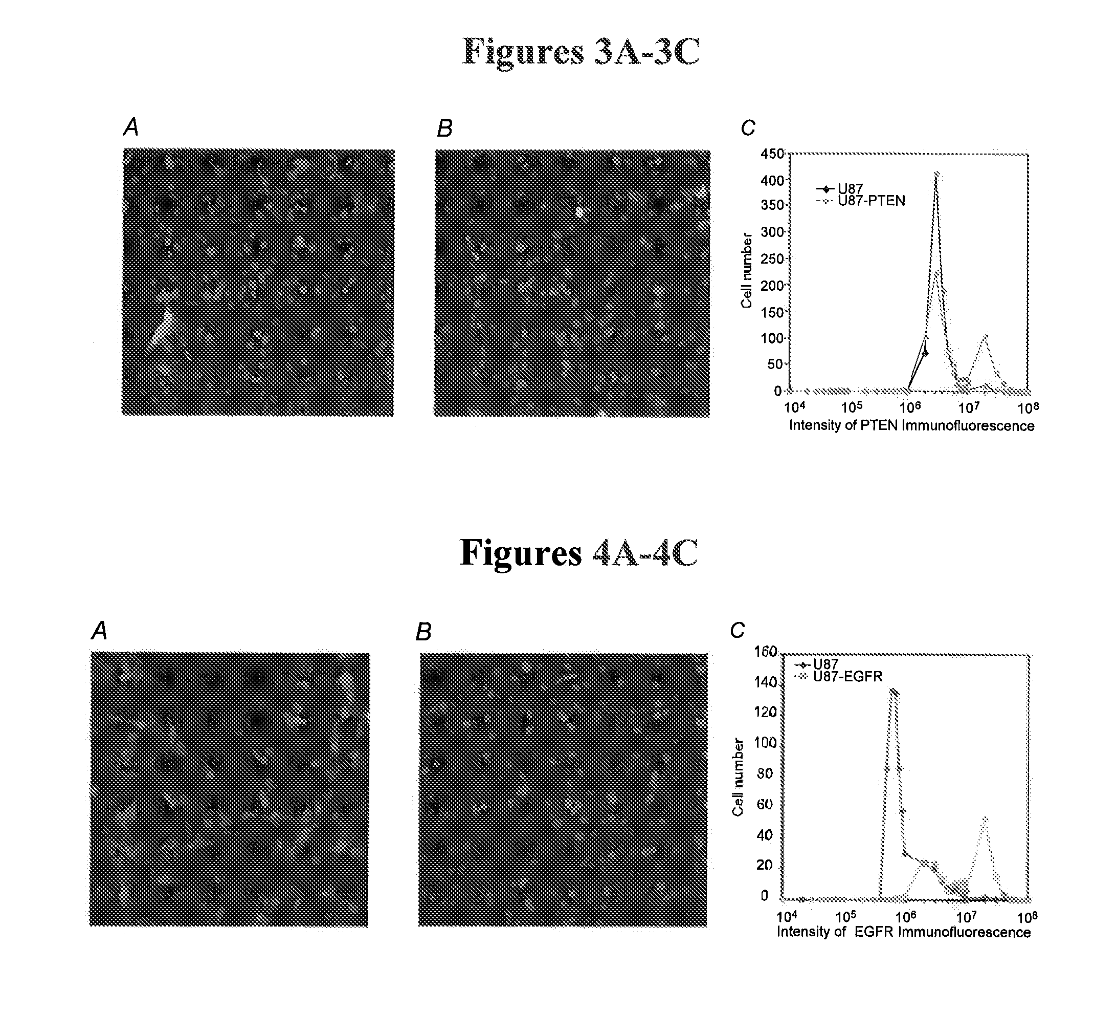
[0105]Data obtained from methods according to some embodiments of the current invention may be in the form, for example, of 2-D or 3-D dot plots (X axis—intensity of one signaling node (in this case, EGFRvIII) and Y axis—intensity of the other signaling node (in this case DAPI)) for 3-D dot plots Z axis—intensity of a third signaling node (FIG. 9). In another embodiment (FIG. 10), the data may be in the form of histograms (X axis—intensity of one signaling node (in this case, EGFRvIII) and Y axis—cell number).
Protocol for Cell Culture for Glioblastoma in a Chip
Materials:
[0106]24-channel poly-L-Lysine-coated cell culture chips[0107]Cell culture medium: 500 mL Dulbecco's Modified Eagle Medium[0108]50 mL Fetal Bovine Serum[0109]5 mL Pen-strep / L-glutamine
Cell Lines and Isogenetic Cells:
[0110]U87[0111]U87-PTEN[0112]U87-EGFR[0113]U87-EGFRvIII[0114]U87-EGFRvIII / PTEN
Automated Pipettes:
[0115]Multichannel (From Matrix Technologies Corporation, two suggested pipettes with Item N...
example c
The Current Model of Pathology
[0284]The current model of pathology diagnosis for cancer is based on the microscopic resemblance of cancer cells to their presumed cell of origin or its developmental precursor. Based on tissue morphological appearance, as well as the presence or absence of a few protein markers, the pathologist concludes a broad pathological diagnosis conveying tumor type and grade. Typically, the patient is treated with relatively toxic, non-specific therapies such as DNA damaging agents and radiation. While this classification and affiliated grading system has proven to be useful for predicting the overall survival for groups of patients and for communicating broad information about the disease category24-29, relatively limited insight is gained about the underlying molecular pathway lesions30. Furthemore, clinically relevant subsets that may differ significantly in their time course and responses to therapy cannot be monitored with the current classification system...
PUM
| Property | Measurement | Unit |
|---|---|---|
| volume | aaaaa | aaaaa |
| volume | aaaaa | aaaaa |
| volume | aaaaa | aaaaa |
Abstract
Description
Claims
Application Information
 - R&D
- Intellectual Property
- Life Sciences
- Materials
- Tech Scout
- Unparalleled Data Quality
- Higher Quality Content
- 60% Fewer Hallucinations
Browse by: Latest US Patents, China's latest patents, Technical Efficacy Thesaurus, Application Domain, Technology Topic, Popular Technical Reports.
© 2025 PatSnap. All rights reserved.Legal|Privacy policy|Modern Slavery Act Transparency Statement|Sitemap|About US| Contact US: help@patsnap.com