
Multipurpose analysis using second harmonic generating nanoprobes

a nanoprobe and second harmonic technology, applied in material analysis, biochemistry apparatus and processes, instruments, etc., can solve the problems of increasing the size of the device, the difficulty of identifying cells or probe molecules, and the mechanism and dynamics of biological processes, so as to improve the electric field

Active Publication Date: 2010-09-16
CALIFORNIA INST OF TECH
View PDF16 Cites 13 Cited by
  • Summary
  • Abstract
  • Description
  • Claims
  • Application Information

AI Technical Summary

Benefits of technology

The current invention is about a new technique for imaging and detecting using nanoprobes that generate second harmonic signals. These nanoprobes have several advantages, including the ability to emit coherent waves without bleaching or saturation. The probes are made up of two different types of nanostructures: an exciter nanostructure and a second probe nanostructure. The exciter nanostructure enhances the electric field within a few nanometers of its vicinity, which can couple with neighboring nanoprobes and be used as a sensitive distance gauge. The second probe nanostructure generates the second harmonic signals. The invention also includes a method for imaging and detecting biological processes and structures with high sensitivity and resolution using a field resonance enhanced second harmonic (FRESH) technique. Additionally, the invention includes a voltage-sensitive SHG nanoprobe that can detect and evaluate the electric field generated by a target species or device.

Problems solved by technology

One of the grand open challenges in modern science is to image or monitor processes or structures that are very small and operate at a fast time-scale.
For example, it is extremely difficult to identify cells or probe molecules and understand the mechanism and dynamics of biological processes at the molecular level with high sensitivity and spatiotemporal resolution, and particularly inside living cells and tissue or liquid.
In addition, the continued miniaturization of integrated circuits has led to increasingly small devices.
These larger electric fields carry the added risk that an electrical short or breakdown effect will occur somewhere in the circuit.
If a circuit does not perform to specification, locating troublesome areas can be stymied by the complexity of modern-day circuits; checking each node of the circuit is time-consuming and technically difficult.
However, these techniques require high costs and some fundamental technological barriers hinder their widespread use.
However, the ultimate need of characterizing biological targets is largely unmet due to fundamental deficiencies associated with the use of fluorescent agents.
For example, fluorescent probes face two major limitations that have a significant impact on the signal strength: 1) dye saturation, because the number of photons emitted by the fluorophore in a given time is restricted by the excited state lifetime, and 2) dye bleaching, which limits the total number of photons per dye.
Among these problems, bleaching is perhaps the most significant issue in the application of fluorescent probes, limiting significantly the length of time that biological targets can be studied.
In addition, autofluorescence from tissue organic components due to illumination absorption can severely limit the signal-to-noise ratio.
Finally, fluorescence is fundamentally an optically incoherent process, and as a result extracting 3D information from the source is inherently difficult.

Method used


Image

  • Multipurpose analysis using second harmonic generating nanoprobes
  • Multipurpose analysis using second harmonic generating nanoprobes
  • Multipurpose analysis using second harmonic generating nanoprobes

Examples


example 1

SHG Nanoprobe Emission Profiling

[0055]FIGS. 1b to 1d provide exemplary data graph for a second harmonic emission profile generated from BaTiO3-nanocrystals in accordance with the current invention, and in comparison to the emission characteristics of conventional. Quantum Dots (QD) (d). As shown, the second harmonic emission ranges from 380 to 485 nm displaying, unlike many other optical probes, discrete emission peaks of around 10 nm, and was generated by conventional two-photon excitation, where the excitation energy ranges from 760 to 970 nm.

[0056]For this data (spectrum, blinking property comparison, and signal saturation comparison) SHG nanomaterial solutions were ultrasonicated, nanofiltered and immobilized in 20% polyacrylamide gel. The spectrum was analyzed with the Meta detector of the Zeiss 510NLO microscope setup. For the blinking property comparison between BaTiO3 and water-soluble QD, both immobilized nanomaterials were imaged 300 times (20 frames / second) using a 40× / 1....

example 2

SHG Nanoprobe Imaging

[0062]To test whether SHG nanoprobes can be detected in live vertebrate tissue, BaTiO3 nanoparticles were injected into one-cell stage zebrafish embryos and imaged. More specifically, in this example the SHG nanocrystal probes of the current invention, BaTiO3 nanocrystals were injected into zebrafish embryos. Several days after cytoplasmic injection (around 72 hpf) excitation of nanocrystals with femtosecond pulsed 820 nm light results in strong SHG signal detectable in epi-direction as well as in trans-direction throughout the whole zebrafish body.

[0063]The injected embryos developed indistinguishably from their uninjected counterparts, demonstrating that barium titanate is physiologically inert and non-toxic to the sensitive zebrafish embryonic cells. Bright signal could be detected superficially and deep within the tissue. Even in deep tissues and organs, the imaging required only low levels of illumination intensity. The in vivo SHG intensity spectra were id...

example 3

Field Resonance Enhanced Second Harmonic Technique

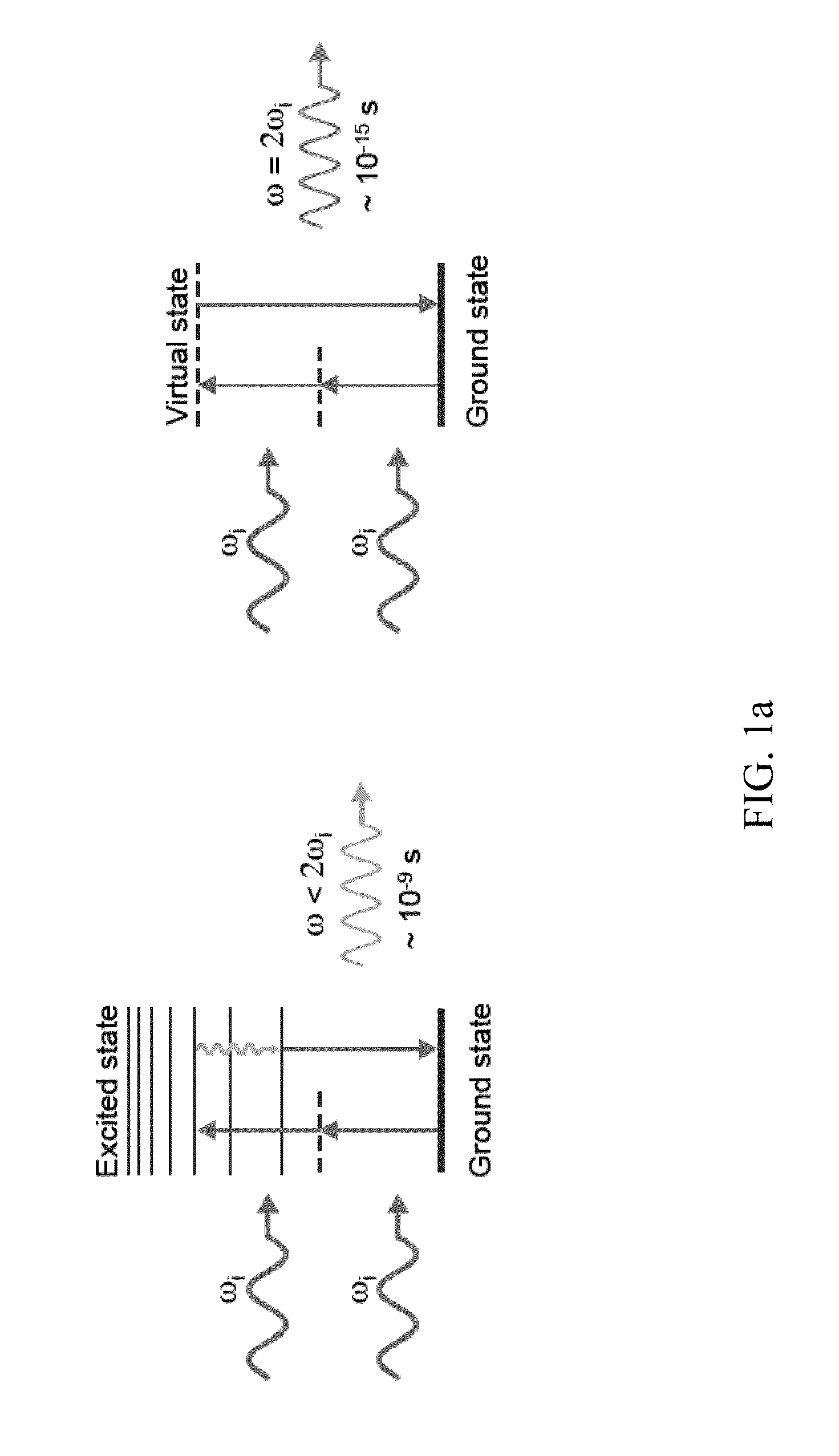
[0066]In addition to simple second harmonic imaging using the second harmonic generating nanoprobes of the current invention, the nanoprobes may also be used in a field resonance enhanced mode to allow access to a number of biological processes that can occur below the nanosecond time frame. Using this field resonance enhanced second harmonic (FRESH) technique in accordance with the current invention it is possible to examine the dynamics of biological processes with high sensitivity and spatiotemporal resolution.

[0067]To understand the potential importance of the FRESH technique it is necessary to examine the inner workings of most biological processes. Besides having highly complex three-dimensional. (3D) structures spanning a large range of length scales, living organisms by their nature are very dynamic: molecular processes such as protein, DNA, and RNA conformations—that take place in a timescale ranging from 100 fs to 100 s whi...


PUM

PropertyMeasurementUnit
excitation wavelengthaaaaaaaaaa
excitation wavelengthsaaaaaaaaaa

Abstract

Second harmonic nanoprobes for multipurpose imaging of samples and a method of using such probes to monitor nucleotide sequencing in a Multi-SHG Detection Imaging (MSDI) modality and to monitor external electric field using voltage sensitive second harmonic generating (SHG) nanoprobes are provided. The SHG nanoprobes are comprised of various kinds of nanocrystals that do not possess an inversion symmetry and therefore are capable of generating second harmonic signals that can then be detected by conventional two-photon microscopy for in vivo imaging of biological processes and structures such as cell signaling, neuroimaging, protein conformation probing, DNA conformation probing, gene transcription, virus infection and replication in cells, protein dynamics, tumor imaging and cancer therapy evaluation and diagnosis as well as quantification in optical imaging for a wide-range of biological and non-biological processes and devices.

Description

CROSS REFERENCE TO RELATED APPLICATIONS[0001]The current application claims priority to U.S. Provisional. Application Nos. 61 / 205,488, filed Jan. 21, 2009 and 61 / 206,960, filed Feb. 6, 2009, the disclosures of which are incorporated herein by reference.STATEMENT OF FEDERAL SUPPORT[0002]The U.S. Government has certain rights in this invention pursuant to Grant Nos. Grant No(s). HD043897 and HGD04071 awarded by the National Institutes of Health.FIELD OF THE INVENTION[0003]The current invention is directed second harmonic generating nanoprobes and methodologies of imaging and analyzing a variety of structures and processes using second harmonic generating nanoprobes.BACKGROUND OF THE INVENTION[0004]One of the grand open challenges in modern science is to image or monitor processes or structures that are very small and operate at a fast time-scale. For example, it is extremely difficult to identify cells or probe molecules and understand the mechanism and dynamics of biological processe...

Claims


Application Information

Patent Timeline
Patent Type & Authority Applications(United States)
IPC IPC(8): G01N33/48G02F1/37
CPCB82Y15/00B82Y20/00C12Q1/6818G01N21/6428G01N21/6458Y10T436/143333G02F1/37G02F2202/36G01N2021/6441C12Q2563/155C12Q2535/101G01N21/636
Inventor PANTAZIS, PERIKLISMASMANIDIS, SOTIRIOSFRASER, SCOTT E.
Owner CALIFORNIA INST OF TECH
Learn More