
Method for induction/differentiation into photoreceptor cell

Inactive Publication Date: 2010-04-29
RIKEN
View PDF4 Cites 47 Cited by
  • Summary
  • Abstract
  • Description
  • Claims
  • Application Information

AI Technical Summary

Benefits of technology

[0010]Use of a method of the present invention makes it possible to efficiently generate retinal progenitor cells or photoreceptors from ES cells without co-culture with retinal tissue. In particular, a method of the present invention is advantageous in that retinal progenitor cells or photoreceptors can be efficiently produced from primate ES cells under defined culture conditions.
[0011]The present invention can largely promote the development of transplantation therapies for retinal diseases based on human ES cells.

Problems solved by technology

By contrast, the in vitro generation of photoreceptors from ES cell-derived progenitor cells remains inefficient unless the progenitor cells are co-cultured with developing retinal tissue.
However, no method has been established for producing photoreceptors under culture conditions without the co-culture.

Method used


Image

  • Method for induction/differentiation into photoreceptor cell
  • Method for induction/differentiation into photoreceptor cell
  • Method for induction/differentiation into photoreceptor cell

Examples


example 1

Methods

Cell Culture

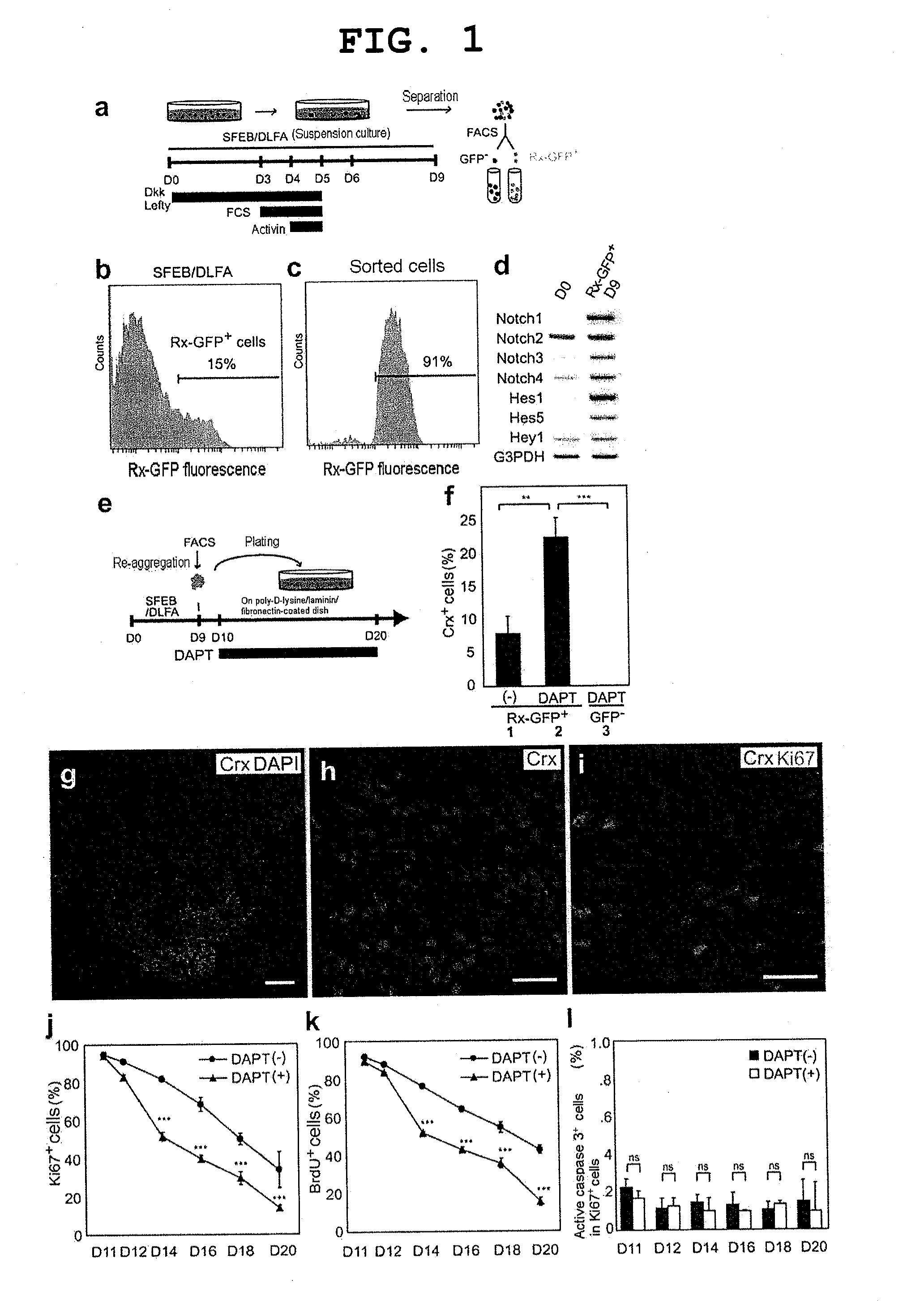
[0140]Methods for mouse ES cell maintenance and differentiation in SFEB conditions were previously reported (reference documents A7, 20). For the sake of the SFEB / DLFA, Dkk1 (R & D Systems, 100 ng / ml, during days 0-5), LeftyA (R & D Systems, 500 ng / ml, during days 0-5), 5% FCS (JRH Biosciences, during days 3-5), and Activin-A (R & D Systems, 10 ng / ml, during days 4-5) were added to a differentiation medium (G-MEM, 5% KSR, 0.1 mM non-essential amino acids, 1 mM pyruvic acid, and 0.1 mM 2-mercaptoethanol) (reference document A6).

[0141]For FACS (see below), until day 9, cell aggregates were incubated in a differentiation medium under suspension culture conditions. After FACS, 1−2×10⁴ sorted cells were re-suspended in a differentiation medium containing 10% FCS, and to produce re-aggregated pellets, the suspension was centrifuged at 800 g for 10 minutes. After cultivation at 37° C. for 1 hour, three to five re-aggregated pellets per cm2 were replated on a poly-D-lysin...

example 2

Methods

Maintenance of Undifferentiated Monkey ES Cells

[0158]Two independent cell lines of cynomolgus monkey ES cells (CMK6 and CMK9) were maintained as described previously (reference documents B26, 28, 29). In summary, undifferentiated ES cells were maintained on a feeder layer of mitomycin C-treated mouse embryonic fibroblasts (STO cells). The STO cells were incubated with 10 μg / mL mitomycin C (Wako, Osaka, Japan) for 2 hours and plated on gelatin-coated dishes. The undifferentiated monkey ES cells were incubated in a DMEM / F-12 (Sigma, St. Louis, Mo.) supplemented with 0.1 mM 2-mercaptoethanol (Sigma), 0.1 mM non-essential amino acids (Sigma), 2 mM L-glutamine (Sigma), 20% knockout serum replacement (KSR; Lot.1139720 and 1219101, GIBCO), 1,000 units / ml leukemia inhibitor (ESGRO; Chemicon, Temecula, Calif.) and 4 ng / ml basic fibroblast growth factor (Upstate Biotechnology, Lake Placid, N.Y.). ES cells were passaged every 3 days using 0.25% trypsin (GIBCO) in PBS supplemented with 1...

example 3

[0180]Mouse ES cells were cultured in the presence of SB-431542 (0.1 to 10 μM) and / or CKI-7 (1 to 100 μM) under SFEB conditions. Five days after the start of the culture, the cells were recovered and re-suspended in a fresh differentiation medium, and, to produce re-aggregated pellets, the suspension was centrifuged at 800 g for 10 minutes. After cultivation at 37° C. for 1 hour, three to five re-aggregated pellets per cm2 were replated onto poly-D-lysine / laminin / fibronectin-coated culture slides, along with differentiation medium. After cultivation for 5 days, the cells were fixed and stained with anti-βII tubulin antibody, and the ratio of βII tubulin-positive cells was determined under microscopy. The other experimental conditions were the same as those for Example 1.

[0181]As a result, with the addition of SB-431542 or CKI-7, the number of βII tubulin-positive cells increased (FIG. 10). This shows that SB-431542 and CKI-7 are each capable of enhancing differentiation of neuronal ...


PUM


Abstract

The present invention provides a method of producing primate retinal progenitor cells, comprising culturing primate embryonic stem cells as suspended aggregates in a serum-free medium, and obtaining retinal progenitor cells from the culture. The present invention further provides a method of producing photoreceptor precursor cells, comprising culturing isolated retinal progenitor cells differentiated from embryonic stem cells, under adhesive conditions, in the presence of a gamma secretase inhibitor, and obtaining a photoreceptor precursor from the culture.

Description

TECHNICAL FIELD[0001]The present invention relates to a method of producing retinal progenitor cells, photoreceptor precursors, photoreceptors and the like.BACKGROUND ART[0002]The number of patients suffering blindness due to a retinal degenerative disease such as age-related macular degeneration or retinitis pigmentosa has been increasing. Because a disorder of photoreceptors is the direct cause of blindness in these diseases, production of photoreceptors (or photoreceptor precursors) in vitro would possibly contribute significantly to research into these diseases and development of therapies for these diseases.[0003]Including the present inventors, many groups have attempted to generate photoreceptors from iris tissue (non-patent documents 1, 2), ciliary tissue (non-patent document 3), or embryonic stem (ES) cells (non-patent documents 4-6). Compared with tissue stem cells, ES cells have the capability of proliferating infinitely and permit production of sufficient numbers of cell...

Claims


Application Information

Patent Timeline
IPC IPC(8): C12N5/0735C12N5/071C12N5/0793C12N5/0797
CPCA61K35/12C12N5/062C12N5/0621C12N5/0623C12N2500/33C12N2500/99C12N2506/02C12N2501/115C12N2501/16C12N2501/385C12N2501/415C12N2501/999C12N2501/113C12N2500/90C12N2533/00C12N2506/08
Inventor TAKAHASHI, MASAYOOSAKADA, FUMITAKAMANDAI, MICHIKOIKEDA, HANAKO
Owner RIKEN
Learn More