Sensitive and rapid detection of viral particles in early viral infection by laser tweezers

Inactive Publication Date: 2010-04-22
KENT STATE UNIV
View PDF20 Cites 16 Cited by
  • Summary
  • Abstract
  • Description
  • Claims
  • Application Information

AI Technical Summary

Benefits of technology

[0002]In the case of viral infection, the infection starts with an initial peak of what is called “viremia” a few days after infection. Viremia is the medical condition where viruses enter the bloodstream and hence have access to the rest of the body. Viremia is followed by the decrease of the viral level as a result of the activation of a person's immune system. The immune system interacts with the virus to either completely control viral infection, partially control viral infection, or not control viral infection at all which leads to increase in virus levels. The initial peak of viremia generally occurs within one or two weeks after infection. This initial peak of virus levels gives medical professionals a window for possible detection of viral copies and rapid diagnosis of viral diseases.
[0003]With viruses, bacterium or other microbes or pathogens, an antibody is a molecule produced by the immune system of a human or an animal in response to a foreign particle or pathogen. The antibody is able to chemically bond to a particular portion of the foreign particle known as the antigen. A foreign particle may have several antigens, though a particular antibody binds to only one of them. The recognition and subsequent binding of the antibody are among the initial stages in the immune response, and specific antibodies are produced by the body in response to particular pathogens. Therefore, the presence of a particular antibody in the blood is an indicator of a particular infection, which may be found before the onset of any signs or symptoms of the disease. In general, it has been the approach to look for the presence of the antibody as an indicator of disease rather than to detect the pathogen directly as the antibody levels in the blood generally far exceed the pathogen levels. After viral infection for example, a person's immune system will start to produce antiviral antibodies. Viral specific antibodies are usually generated within several weeks after infection. A medical professional can accurately diagnose a patient with a possible viral infection by examining the interaction of the anti-viral antibodies. However, levels of antibody may only become detectable using known techniques when the controllable stage of the viral disease already passes.
[0006]Early treatment of an infectious disease, such as that caused by a virus, bacterium or other pathogen, can provide an efficient therapy and immediate control of a virus (or other microbe) outbreak. A rapid and accurate diagnosis of infection at the early stage of infection provides many benefits. There are currently three categories of diagnostic methods: microscopic diagnosis, molecular diagnosis, and serologic diagnosis (which aims to detect the level of antibody against specific pathogens such as virus and microbes as described above). In microscopic diagnosis, individual viral particles can be observed with electron microscopes. The microscopic detection of organisms stained with fluorescent dyes or other markers attached to antibodies has been developed for the specific identification of some viruses. Although useful, fluorescent dyes will be destroyed under prolonged exposure of the excitation light (where absorption is maximal) due to photoactivated chemical reactions, causing bleaching, and inhibiting proper detection. Such a problem makes the use of fluorescent dyes problematic in attempting to detect small numbers of or single virus particles, or other pathogens or microbes. Since only a few molecules of fluorescent dye can be attached to the single virus, which is usually very small (hundreds of nanometers and below), proper detection is inhibited. Due to the small amount of the dyes, bleaching only a few of them can cause the dramatic decrease of the signal and even total loss of the signal.
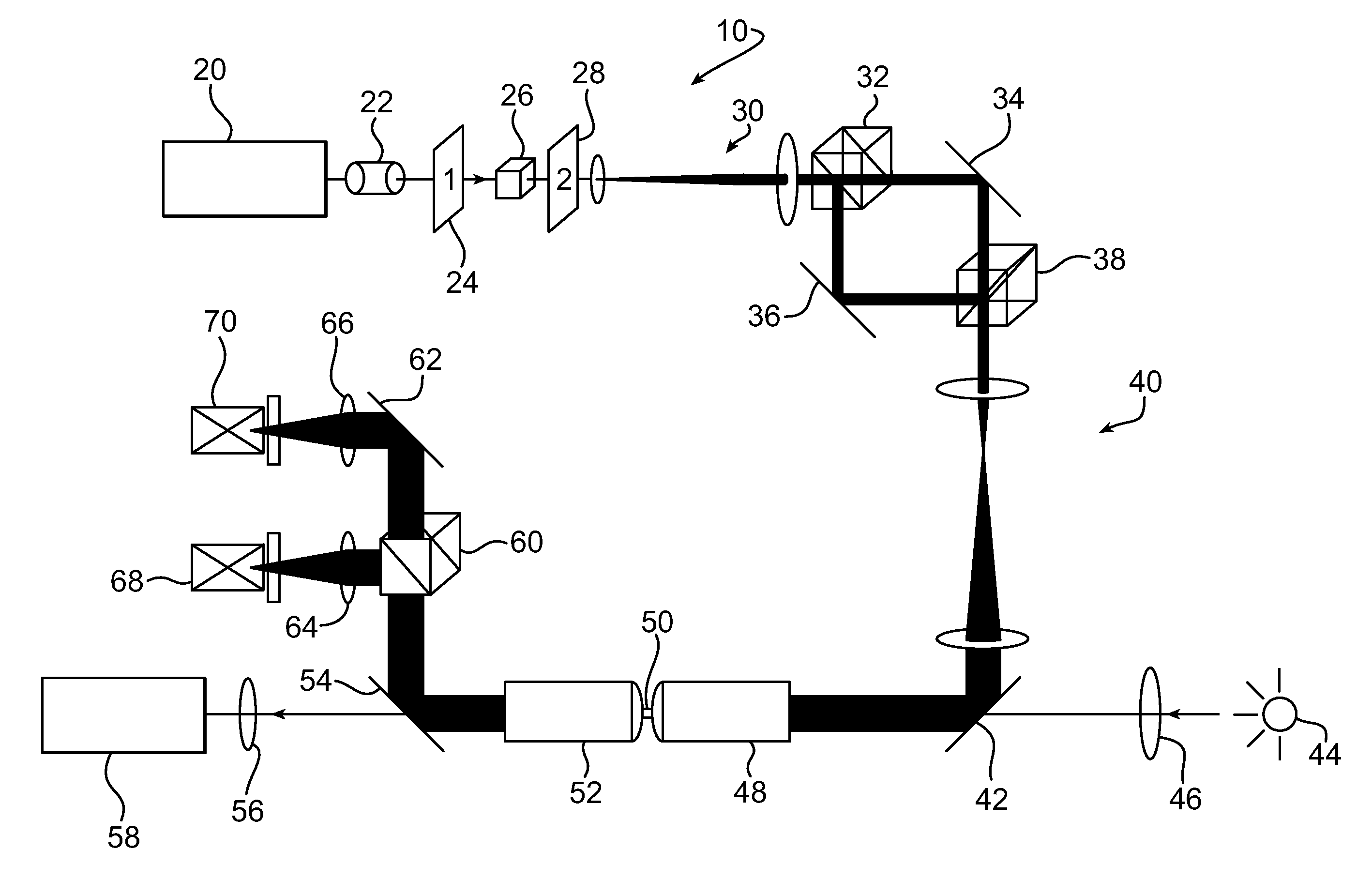
[0009]The present disclosure relates to systems and methods that allow for highly sensitive and simple detection method of viral and other microbe particles during the initial peak of viremia or infection. The system and methods may utilize optical tweezers to provide a rapid and accurate detection method to identify the infection. By using focused laser beams, optical tweezers can trap and remotely manipulate dielectric particles. The particles may include cells, bacterial and viral particles. The embodiments of the apparatus and methods of the present invention addresses the deficiencies of prior systems and methods, and allow a small reaction substrate formed at least one nano- or micro-particle to be used in detecting as little as a single microbe with high sensitivity. The systems and methods provide for detection of a microbe or other analyte which can be effectively used in various environments, including where the virus, bacterium or the like is released, such as for combating bioterrorism in field environments, as in the case of bioterrorism for example. The invention simplifies use and detection at a very early stage of infection, and can be used to detect multiple analytes or microbes.

Problems solved by technology

However, levels of antibody may only become detectable using known techniques when the controllable stage of the viral disease already passes.
Although useful, fluorescent dyes will be destroyed under prolonged exposure of the excitation light (where absorption is maximal) due to photoactivated chemical reactions, causing bleaching, and inhibiting proper detection.
Such a problem makes the use of fluorescent dyes problematic in attempting to detect small numbers of or single virus particles, or other pathogens or microbes.
Due to the small amount of the dyes, bleaching only a few of them can cause the dramatic decrease of the signal and even total loss of the signal.
This is especially true when pathogens are highly contagious and no treatment is available for those potentially employed by bioterrorists.
At this stage, it is often too late to prevent the spread of infection or an outbreak in the population.

Method used


Image

  • Sensitive and rapid detection of viral particles in early viral infection by laser tweezers
  • Sensitive and rapid detection of viral particles in early viral infection by laser tweezers
  • Sensitive and rapid detection of viral particles in early viral infection by laser tweezers

Examples


example 2

[0027]The second example according to the presently disclosed system and method may be used as a control sample. The second sample may be made by coating the plurality of microspheres 100 with a protein binding agent 140 such as streptavidin. Next, the protein coated microspheres 100 are mixed with a control antibody. In the present embodiment, the control antibody is a buffer solution containing rabbit inmunoglobulin G (IgG) control antibody. The second sample may be incubated for 20 to 30 minutes at room temperature and then centrifuged. After centrifuging, the sample is mixed with a buffer solution and mixed thoroughly.

[0028]According to the presently disclosed system and method, a solution containing the analyte 130 to be detected, such as an antigen or a virus, may be prepared or acquired from environmental or human medium, for example, water, or human blood. In the present embodiment, the anlayte is an HIV virus or a virus like particle (VLP, Functional Genetics, Inc.). The vi...


PUM


Abstract

The present system and methods allow for low level detection of as little as single pathogen particles, such as viral or bacterial particles, during the early stage of infection. An optical trapping system, such as laser tweezers, are used to trap a substrate to which an analyte has been bound to detect and record the thermal motion of an antibody-antigen interaction that may occur between an anti-viral antibody-coated microsphere and a viral particle for example. The system may be equipped with a detection system such as a position sensitive photodetector (PSD) to record the thermal motion of a trapped microsphere and particle at a certain frequency. The thermal motion data may be Fourier transformed into a power spectrum, which may be transformed into an output value using a Lorentzian equation. The power spectrum of the trapped microsphere may be recorded before and after binding of the pathogenic particle to determine the presence thereof.

Description

BACKGROUND AND SUMMARY[0001]The present disclosure relates to a system and method for detecting analytes in a medium and more particularly to method for detecting low levels of analytes such as antigens including viruses or other microbes and pathogens.[0002]In the case of viral infection, the infection starts with an initial peak of what is called “viremia” a few days after infection. Viremia is the medical condition where viruses enter the bloodstream and hence have access to the rest of the body. Viremia is followed by the decrease of the viral level as a result of the activation of a person's immune system. The immune system interacts with the virus to either completely control viral infection, partially control viral infection, or not control viral infection at all which leads to increase in virus levels. The initial peak of viremia generally occurs within one or two weeks after infection. This initial peak of virus levels gives medical professionals a window for possible detec...

Claims


Application Information

Patent Timeline
IPC IPC(8): C12Q1/70C12M1/00
CPCG01N33/54313G01N33/56983G01N33/54373
Inventor MAO, HANBINMAO, HANWENMAO, ZIANMAO, JIANGSEN
Owner KENT STATE UNIV
Why Eureka
Try Eureka