
Biomaterial for Suturing

a technology of biomaterials and sutures, applied in the field of biomaterials for suction, can solve the problems of suture closure, particularly delicate sutures, and generating areas where fluids can escap

Inactive Publication Date: 2009-11-26
AUTONOMOUS UNIVERSITY OF MADRID +1
View PDF20 Cites 22 Cited by
  • Summary
  • Abstract
  • Description
  • Claims
  • Application Information

AI Technical Summary

Benefits of technology

[0018]The biomaterial for suturing referred to in this invention is composed of a physiologically compatible material known as support material which is coated with a cellular population with proliferative and / or differentiation capacity, characteristics which are necessary to participate in the regeneration of the sutured tissue. Thus, this biomaterial for suturing is not limited to bringing the two ends of the open wound together, but also contributes actively to scar tissue formation, accelerating the tissue repair process. This innovation represents an important advantage, especially in the case of sutured internal organs and particularly for intestinal anastomosis as a result of resectioning the gastrointestinal tract or urogenital area.
[0033]A preferred embodiment refers to the use of progenitor cells from a specialized cellular lineage obtained from the patient's stem cells which express at least one of the characteristics of the specialized progenitor cells as the cellular population used to coat the biomaterial for suturing to which the invention refers. This prevents the generation of inflammatory problems and rejection, as and the components of the suturing material would be concealed from the immunological system by coating it with autologous stem cells, which would undoubtedly improve the tissue repair process.

Problems solved by technology

Any open wound poses the risk of infection and a channel for air and organic fluids to escape, making its closure an urgent necessity.
In some situations; these sutures are particularly delicate due to the healing difficulties of the tissues they are used on.
One of the biggest disadvantages of sutures is the fact that the diameter of the needle is larger than that of the thread, so that the point where the needle is inserted is not taken up completely by the thread, thus generating areas where fluids can escape.
This deficient closure of the wound is frequently the cause of post-operative complications, such as in the case of intestinal anastomosis in patients with carcinoma or diverticulosis who have had part of their intestine removed, after which the two healthy ends are joined together.
In these patients, when the closure is incomplete faecal matter can leak into the surrounding tissues, which is a cause of peritonitis and places the patient's life at risk.
A limiting factor in the use of staples as a method of primary intention healing is the need to have access to the upper and lower part of the tissues being joined together.
Furthermore, the force exerted when inserting the staples may cause the tissue to tear.
The biggest disadvantage of biological adhesives is the risk of viral transmission.
Moreover, there are several disadvantages to the synthetic bioadhesives currently available.
Most of them come in liquid form and are therefore difficult to apply, so they are used primarily on superficial wounds.
They are also allergenic and potentially toxic substances.
It has been described that these materials induce an inflammatory response in the body, which contributes to a delay in the regeneration and healing of the tissue, thus greatly limiting their usefulness (Aronson et al., 1970; Milde et al., 1989).
However, one of the disadvantages is the allergenic nature and potential toxicity of bioadhesives.
The ability to join biological tissues together has been one of the principal challenges of biomedical research.

Method used


Image

  • Biomaterial for Suturing
  • Biomaterial for Suturing
  • Biomaterial for Suturing

Examples


example 1

Suturing Threads Coated with Adult Human Stem Cells from Adipose Tissue

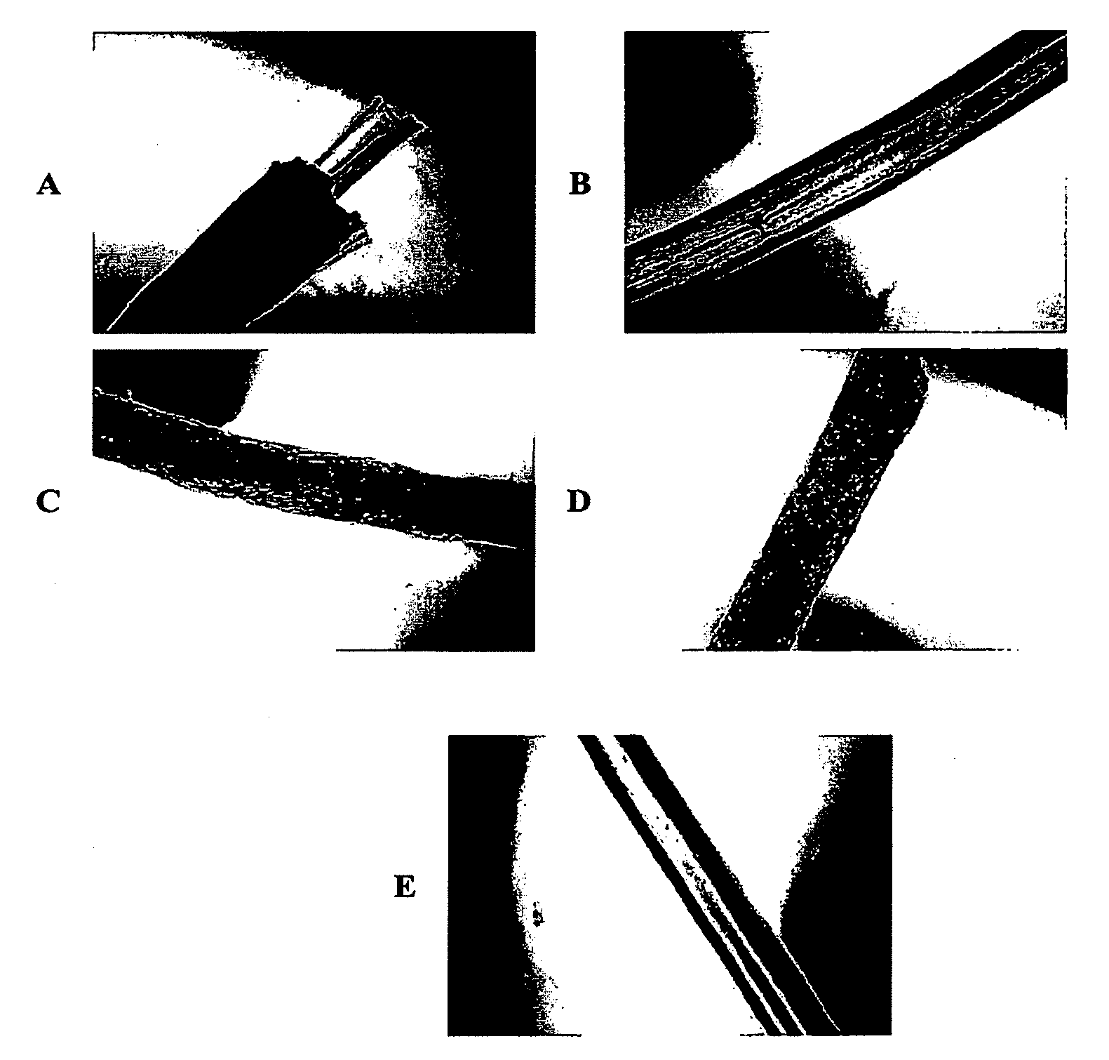
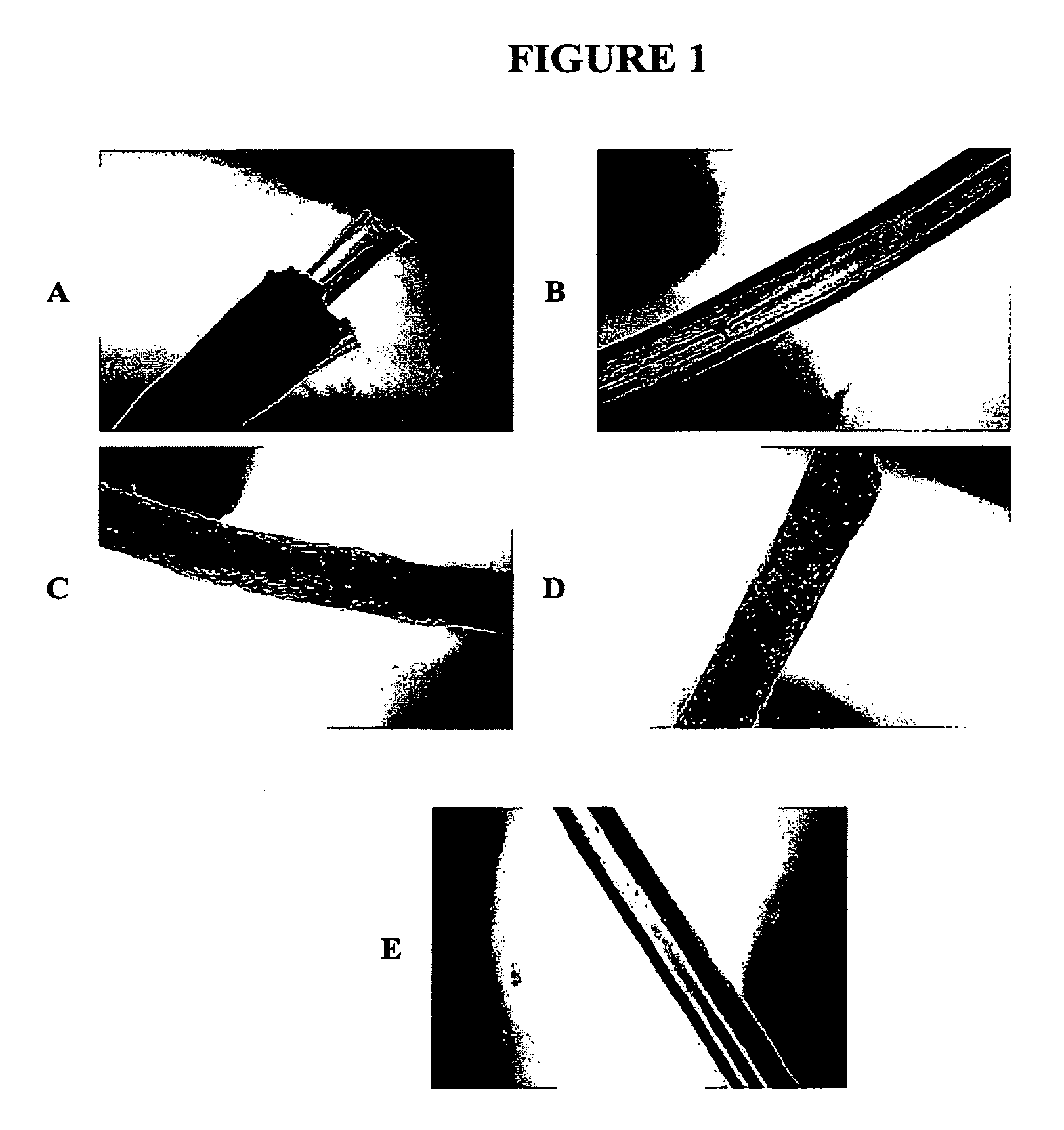
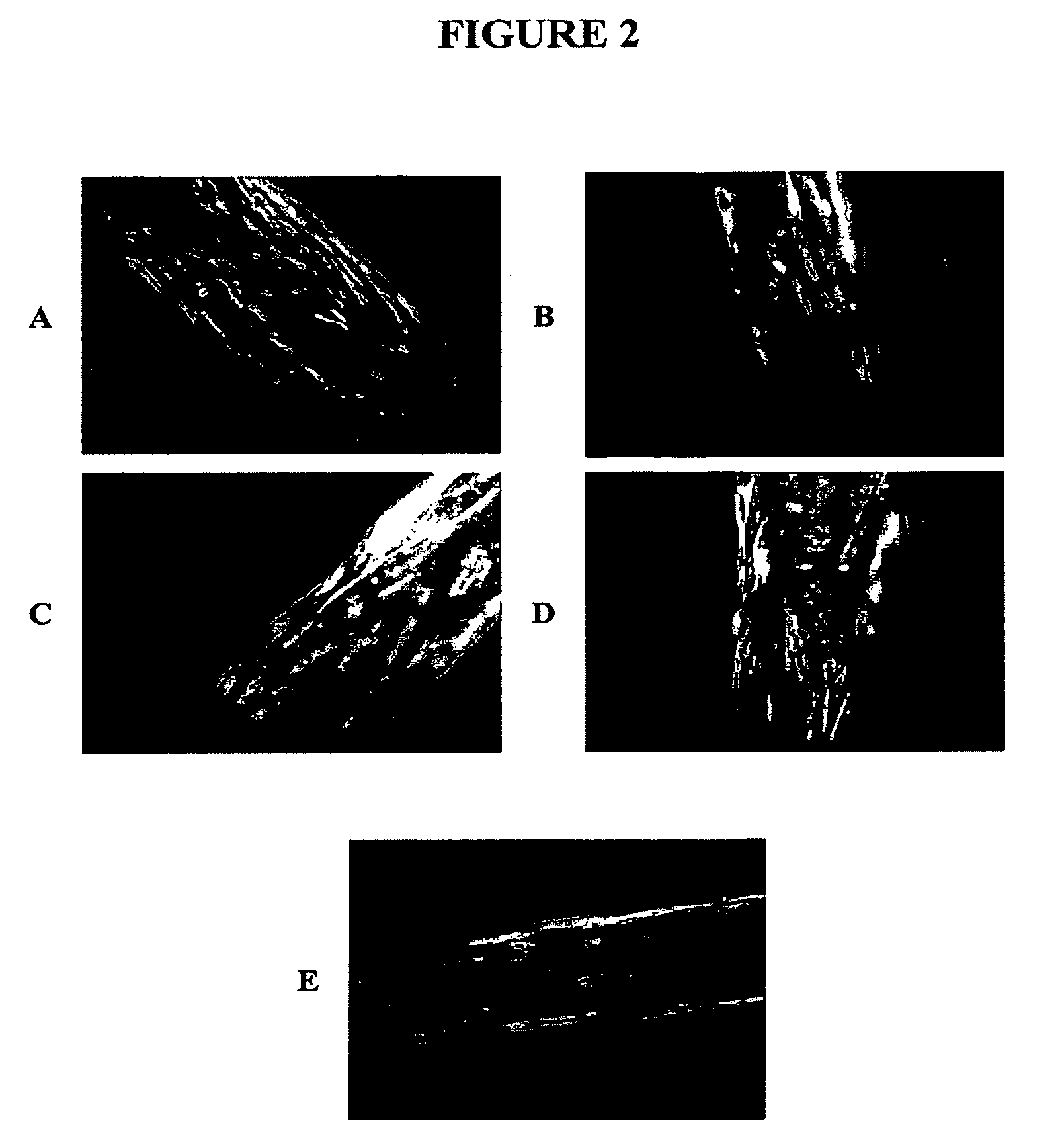
[0064]The object of this experiment was to study the capacity of a certain type of cell to adhere to different types of suturing threads acting as the support material for the biomaterial for suturing of this invention.

[0065]1.1. Materials

[0066]Five different types of suturing thread, all of the same thickness 3-0 (2 Ph.Eur.) were used, all synthetic (Table 1).

[0067]Adherent adult human lipo-derived stem cells (LDSC) were used as the cellular population for coating, after first being transduced with retrovial vectors that code for Cop-GFP, green fluorescent protein, used as a gene marker.

[0068]1.1.1. Isolation of LDSC

[0069]The adipose tissue was obtained by liposuction. A cannula with a blunt end was introduced into the subcutaneous space through a small periumbilical incision (less than 0.5 cm in diameter). The suction was performed by moving the cannula along the adipose tissue compartment located under the abd...

example 2

Use of the Biomaterial for Suturing for Intestinal Anastomosis

[0093]The object of this test was to determine the characteristics of the biomaterial for suturing provided by this invention and the advantages it offers compared to conventional sutures by performing colical anastomosis in rats.

[0094]After performing the anastomosis with the biomaterial for suturing of the invention and simultaneously with non-cell-coated threads used as a negative control, a series of parameters were determined to evaluate the status of the anastomotic lesion and compare the results obtained with both types of thread.

[0095]2.1. Surgical Procedure

[0096]2.1.1. Animals and Suturing Material

[0097]12 adult BDIX rats with weights ranging between 130-260 grams were used in the experiment. Two to obtain the stem cells from the 4 rats from the subdermal adipose tissue and ten to study the colic sutures. The cell-coated threads were prepared following a protocol similar to that illustrated in example 1. The BDIX...


PUM

PropertyMeasurementUnit
sizeaaaaaaaaaa
diameteraaaaaaaaaa
concentrationaaaaaaaaaa

Abstract

A biomaterial for suturing comprising a physiologically compatible support material coated with a cellular population with proliferative and / or differentiation capacity, characteristics which facilitate the regeneration of the sutured tissue. This biomaterial for suturing not only brings together the two edges of an open wound, but also contributes actively to the healing process, thereby accelerating the tissue repair process. Also disclosed are methods for making the biomaterial and methods for using the biomaterials in therapy.

Description

[0001]This invention refers to a material for suturing and its applications. More specifically, the invention refers to a material for suturing covered with cells that contribute in a biologically active way to the tissue repair process and therefore the healing of wounds.BACKGROUND OF THE INVENTION[0002]Any open wound poses the risk of infection and a channel for air and organic fluids to escape, making its closure an urgent necessity. In superficial skin wounds that do not pierce the dermis, spontaneous closure takes place when the edges of the wound come together, while in wounds where there is a clear separation of tissues only surgical action (suturing of the wound) can achieve this primary closure, also known as primary intention healing.[0003]Traditionally, the suture has been the classical method for bringing the edges of the wound together in order to achieve the rapid tissue repair. Primary intention healing by suturing consists of bringing the edges of the wound together ...

Claims


Application Information

Patent Timeline
Patent Type & Authority Applications(United States)
IPC IPC(8): A61L17/06C12N5/06A61B17/04A61B17/064
CPCA61L17/145A61L27/3839A61L27/3804A61L17/00C12N5/0667
Inventor GARCIA OLMO, DAMIANMIGUEL, GEMA FERNANDEZGONZALEZ DE LA PENA, MANUEL A.
Owner AUTONOMOUS UNIVERSITY OF MADRID
Learn More