Primer and Probe for Detection of Mycobacterium Intracellulare, and Method for Detection of Mycobacterium Intracellulare by Using the Primer and Probe

a technology of mycobacterium and probe, which is applied in the field of primer and probe for detection of mycobacterium intracellulare, can solve the problems of difficult to differentiate tuberculosis from non-tuberculous i>mycobacterium, no specific clinical symptoms of non-tuberculous i>mycobacterium, and infectivity to human exert infection, etc., to achieve false diagnosis results, detect and diagnose more quickly, and high accuracy

Inactive Publication Date: 2009-11-05
WAKO PURE CHEMICAL INDUSTRIES
View PDF16 Cites 6 Cited by
  • Summary
  • Abstract
  • Description
  • Claims
  • Application Information

AI Technical Summary

Benefits of technology

[0026]According to the method for detection of M. intracellulare using the primer and / or probe of the present invention, M. intracellulare can be detected and diagnosed more rapidly and with high accuracy compared to the conventional bacterium identification method by a cell culture assay and the like. In addition, by performing the detection using the method of the present invention, any false-positive result in diagnosis can be eliminated as compared with the diagnosis method by PCR using a conventional primer and / or a probe, and as the results, M. intracellulare can be detected and diagnosed with higher accuracy and preciseness in a specific manner. Further, by the use of the detection method of the present invention, M. intracellulare cell can also be quantified.

Problems solved by technology

However, on rare occasions, they may exert infectivity to human.
However, nontuberculous mycobacterium diseases do not show any specific clinical symptom.
Therefore, it is quite difficult to differentiate tuberculosis from nontuberculous mycobacterium disease by clinical observation and histopathological manifestation, and to specify the species of the nontuberculous mycobacterium.
However, by this test, it can be recognized only whether the pathogenic bacterium is “acid-fast bacterium-positive” or not, and cannot be identified whether the pathogenic bacterium is tuberculosis bacteria or nontuberculous mycobacterium.
However, in general, growth of genus Mycobacterium is slow; for example, it takes 3 to 4 weeks only for its isolation culture.
Accordingly, the conventional basic method, in which a diagnostic outcome on whether the disease is tuberculosis or not is obtained by conducting the above described smear examination and a cell culture assay, is a considerably time-consuming method.
However, in the usual PCR method, the number of bacteria cannot be determined.
Furthermore, the method has other problems such as that judgment of false positive tends to be made due to too high sensitivity.
1). However, by this method, discrimination between M. intracellulare and M. avium cannot be ach
ieved. In addition, in the PCR using the employed rps1 primer (a primer designed from the ribosomal protein s1 gene region of M. intracellulare), even when the isolated strain of M. avium is used as a sample, the amplification product has also been detected; there remains a problem in terms of the specificity for M. intra
However, in the PCR using the aforementioned primer, the primer extension product can be obtained not only when the sample is avian tuberculosis bacterium (M. avium) but also M. intracellulare, and therefore, this determination method can not be said as a specific method for M. intracellulare< /
i>. In addition, the method, whereby the discrimination between both bacterial species is carried out based on the chain length of the primer extension product, is cumbersome; and it is conceivable that different determination may be made depending on the judge; and in consequence, the method cannot be said as a reliable determination met
Because of this, MAC can be detected by this method; however, it is impossible to detect M. intracellulare specifically.
In addition, even when MAC is detected, there can be an instance where false-positive result is provided.
However, specific detection of M. intracellulare is impossible to achieve without detection of M. avium.
However, in the LAMP method, there remains some problems such as that the nucleotide sequence of amplified DNA cannot be determined; that efficient length of DNA to be amplified is limited; and that the method provides false-positive result occasionally; and the like.

Method used


Image

  • Primer and Probe for Detection of Mycobacterium Intracellulare, and Method for Detection of Mycobacterium Intracellulare by Using the Primer and Probe
  • Primer and Probe for Detection of Mycobacterium Intracellulare, and Method for Detection of Mycobacterium Intracellulare by Using the Primer and Probe
  • Primer and Probe for Detection of Mycobacterium Intracellulare, and Method for Detection of Mycobacterium Intracellulare by Using the Primer and Probe

Examples


example 1

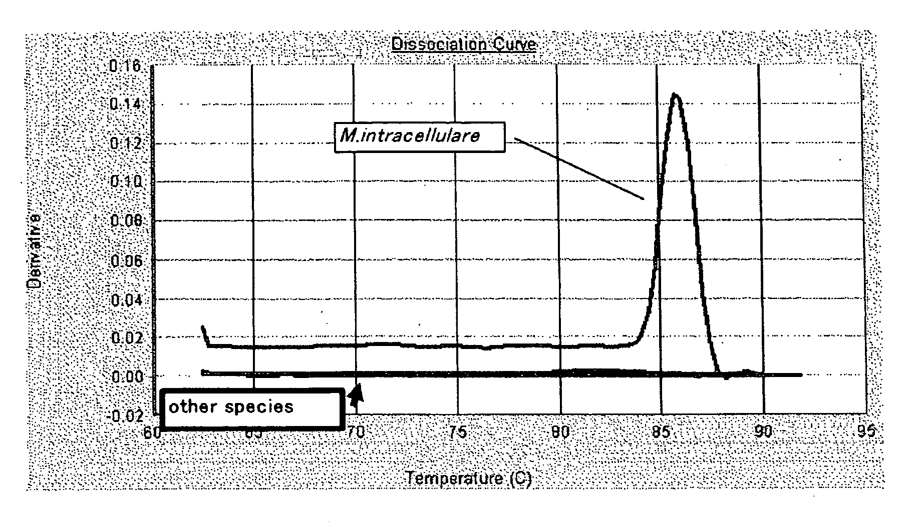
Evaluation on Specificity of the Candidate Clone for M. intracellulare

[0447]For the 8 candidate clones obtained in Experimental Example 1, evaluation experiment through the use of PCR amplification system was performed to investigate the potential use of these clones in the specific detection system for M. intracellulare by using nucleic acid amplification detection system.

(1) Synthesis of the Primer for PCR

[0448]Firstly, based on the result of sequence analysis (nucleotide sequence) of the candidate clone 1, the primer sequence for the PCR amplification detection, namely, the oligonucleotides of “5′-GTTCAGCAGATCGTCGTAGG-37” (SEQ ID NO: 9) and “5′-CTCTTGACGAGGCAAAACAT-3”, (SEQ ID NO: 10) were designed using a primer design tool on the web, Primer 3 (Whitehead Institute for Biomedical Research). Hereinafter, the primer having the nucleotide sequence shown in SEQ ID NO: 9 is referred to as “02_Fw1” and the primer having the nucleotide sequence shown in SEQ ID NO: 10 is referred to as...

example 2

Verification of Sensitivity of the Candidate Clone to Detect M. Intracellulare

[0467](1) Synthesis of the Primer for PCR for Detection of M. intracellulare

[0468]Using the same equipment and by the same procedure as described in (1) of Example 1, the primer 02_Fw1 and the primer 02_Rv1 were synthesized.

(2) Preparation of the Probe for the Detection of M. intracellulare

[0469]From the nucleotide sequence shown in SEQ ID NO: 139 (155 nucleotides) which was anticipated to be amplified by the PCR using 02_Fw1 and 02_Rv1 as primers, a sequence “5′-ATACGTGCCCAGAAGCTCTACCGAGAT-3′” to be used as a probe was designed, and an oligonucleotide consisting of this sequence was synthesized (SEQ ID NO: 204; hereinafter, the oligonucleotide probe having this sequence is described as INT 0 2_F1R1_FAMTAM). The 5′-terminal of this oligonucleotide was labeled with a reporter dye of FAM and the 3′-terminal was labeled with a reporter quencher of TAMRA, and thus a labeled oligonucleotide probe (TaqMan™ Fl...


PUM

PropertyMeasurementUnit
timeaaaaaaaaaa
temperatureaaaaaaaaaa
pressureaaaaaaaaaa

Abstract

The present invention discloses an oligonucleotide which comprises a part or the entire sequence of the nucleotide sequence shown in SEQ ID NO: 1, SEQ ID NO: 2, SEQ ID NO: 3, SEQ ID NO: 4, SEQ ID NO: 5, SEQ ID NO: 6, SEQ ID NO: 7 or SEQ ID NO: 8, or a part or the entire sequence of a sequence complementary to the nucleotide sequence shown in SEQ ID NO: 1, SEQ ID NO: 2, SEQ ID NO: 3, SEQ ID NO: 4, SEQ ID NO: 5, SEQ ID NO: 6, SEQ ID NO: 7 or SEQ ID NO: 8, wherein the oligonucleotide is capable of hybridizing with a nucleotide sequence of Mycobacterium intracellulare gene; a primer or a probe for the detection of M. intracellulare, which comprises the aforementioned oligonucleotide; and a method for detection of M. intracellulare using the aforementioned primer and/or the probe.
According to the detection method of the present invention, any false-positive result in diagnosis can be eliminated and detection or diagnosis of M. intracellulare can be carried out with higher accuracy, more preciseness, and more specifically compared to a conventional diagnostic method employing a cell culture assay or a PCR assay. The method also enables to quantify a microbial cell.

Description

TECHNICAL FIELD[0001]The present invention relates to a method for detecting and / or identifying Mycobacterium intracellulare (hereinafter optionally abbreviated as M. intracellulare) through the use of amplification of nucleic acid and detection system thereof.BACKGROUND ART[0002]Nontuberculous mycobacterium is a gram positive bacillus having acid-fast characteristics classified into genus Mycobacterium (hereinafter, optionally abbreviated simply as M.), and is a kind of acid-fast bacterium other than tuberculosis complex and Mycobacterium leprae. Fifteen to 20% of cases showing positive for sputum smear examination for acid-fast bacterium have been diagnosed thereafter to be nontuberculous mycobacterium by the test for the identification of bacterial species.[0003]Among nontuberculous mycobacterium, clinically problematic bacterial species are blown to be M. intracellulare, Mycobacterium kansasii, Mycobacterium marinum, Mycobacterium gordonae, Mycobacterium szulgai, Mycobacterium a...

Claims


Application Information

Patent Timeline
Patent Type & Authority Applications(United States)
IPC IPC(8): C12Q1/68C07H21/04
CPCC12Q1/689
Inventor ISHIKAWA, TOMOKAZU
Owner WAKO PURE CHEMICAL INDUSTRIES
Why Eureka
Try Eureka