Reagents for the Detection of Protein Phosphorylation in Leukemia Signaling Pathways

a technology of protein phosphorylation and signaling pathway, which is applied in the field of antibodies and peptide reagents for the detection of protein phosphorylation, and to protein phosphorylation in cancer, can solve the problems of anemia, immunodeficiency and coagulation deficiencies, and remains fatal in 80% of treated patients, and is not yet well understood

Inactive Publication Date: 2009-10-22
CELL SIGNALING TECHNOLOGY
View PDF1 Cites 2 Cited by
  • Summary
  • Abstract
  • Description
  • Claims
  • Application Information

AI Technical Summary

Problems solved by technology

Yet, in spite of the importance of protein modification, it is not yet well understood at the molecular level, due to the extraordinary complexity of signaling pathways, and the slow development of technology necessary to unravel it.
The acute forms of the disease rapidly progress, causing the accumulation of immature, functionless cells in the marrow and blood, which in turn results in anemia, immunodeficiency and coagulation deficiencies, respectively.
Without treatment patients rarely survive beyond 6-12 months, and despite continued development of new therapies, it remains fatal in 80% of treated patients (Source: The Leukemia & Lymphoma Society (2004)).
There is, therefore, relatively scarce information about kinase-driven signaling pathways and phosphorylation sites relevant to the different types of leukemia.
This has hampered a complete and accurate understanding of how protein activation within signaling pathways is driving these complex cancers.
However, misdiagnosis can occur since some leukemia cases can be negative for certain markers, and because these markers may not indicate which genes or protein kinases may be deregulated.

Method used


Image

  • Reagents for the Detection of Protein Phosphorylation in Leukemia Signaling Pathways
  • Reagents for the Detection of Protein Phosphorylation in Leukemia Signaling Pathways
  • Reagents for the Detection of Protein Phosphorylation in Leukemia Signaling Pathways

Examples


example 1

Isolation of Phosphotyrosine-Containing Peptides from Extracts of Leukemia Cell Lines and Identification of Novel Phosphorylation Sites

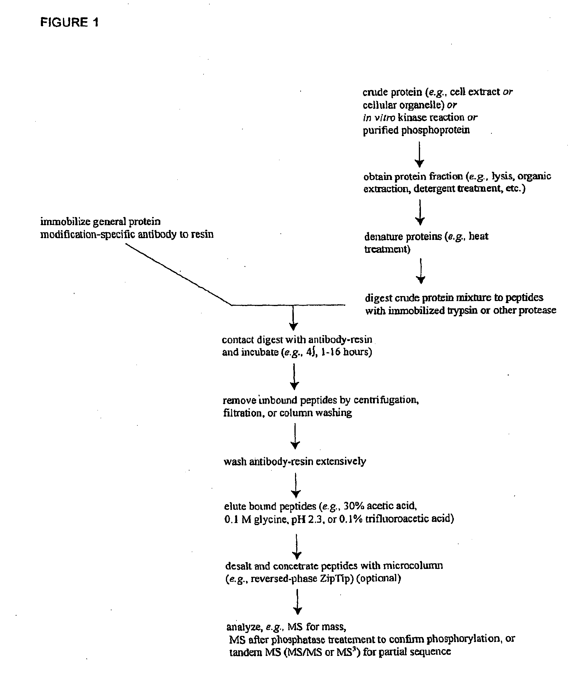
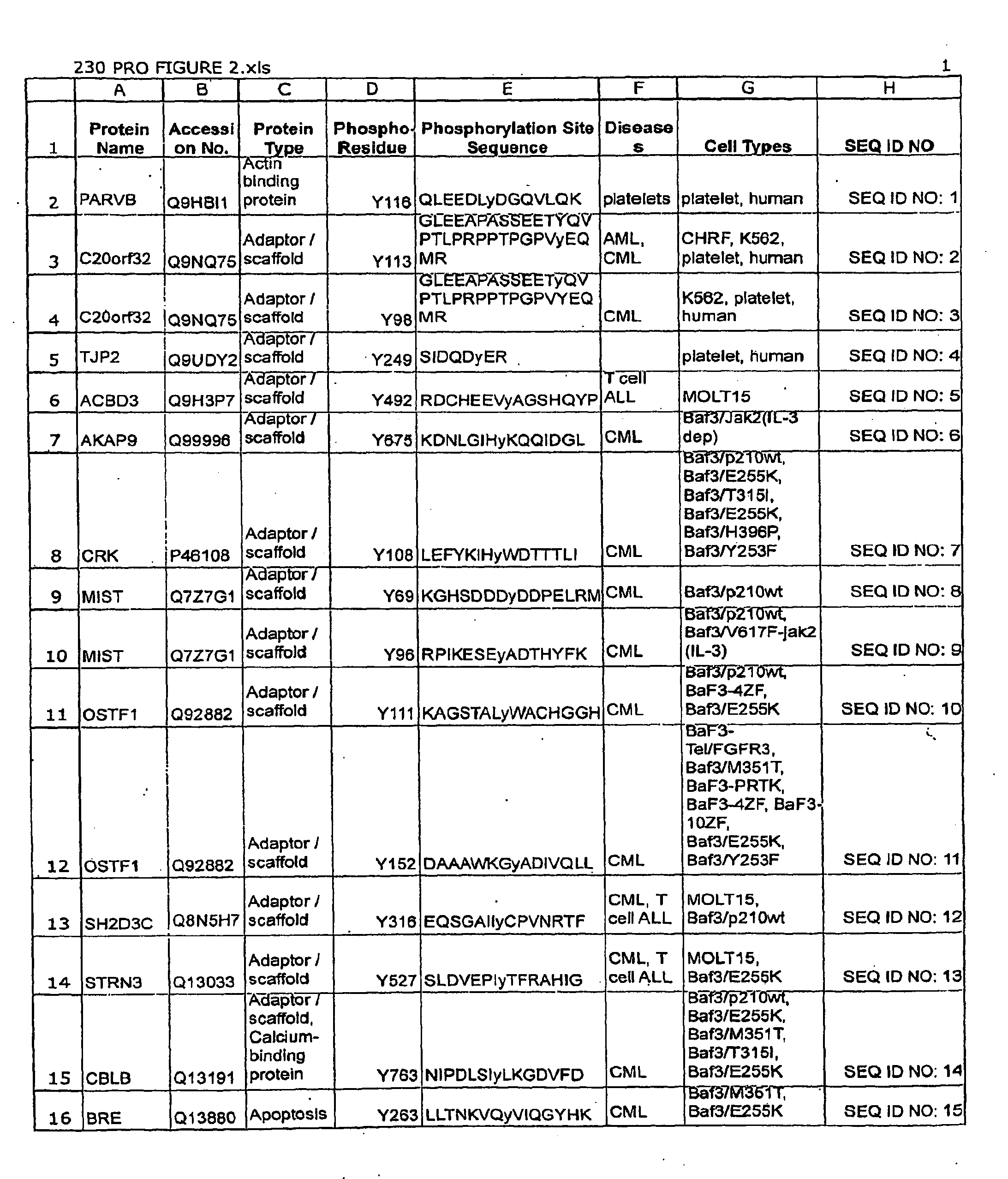
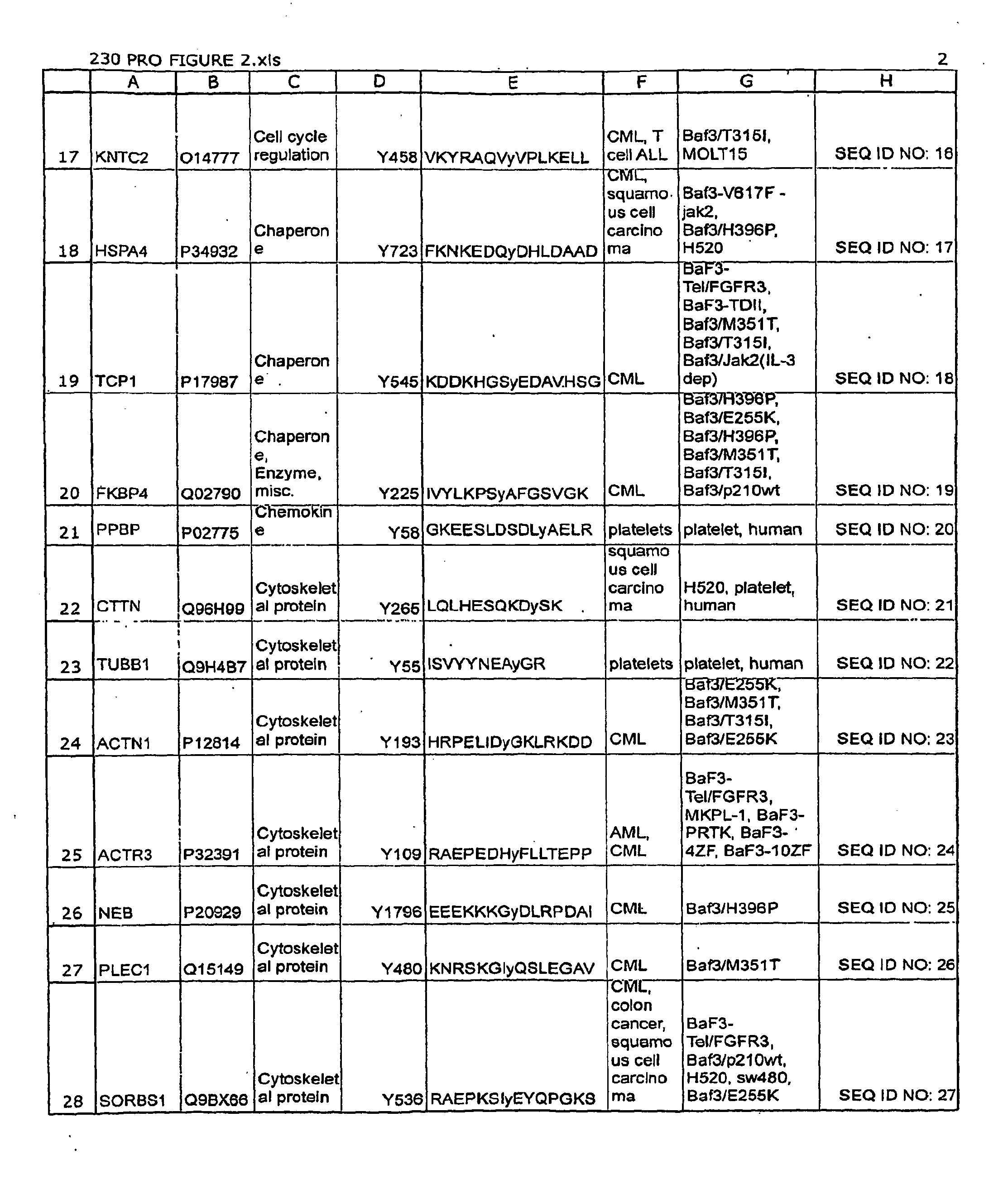
[0129]In order to discover previously unknown Leukemia-related signal transduction protein phosphorylation sites, IAP isolation techniques were employed to identify phosphotyrosine- and / or phosphoserine-containing peptides in cell extracts from the following human Leukemia cell lines and patient cell lines: human platelets, human umbilical vein endothelial cells, K562 (human CML), CMK (human AML), MOLT15 (human ALL), MKPL-1 (human AML), Molm14 (human AML), CHRF (human AML), H520 (human non-small cell lung carcinoma), SW480 (human colorectal carcinoma), OPM-1 (human multiple myeloma), UT-7 (human AML), H3255 (human non-small cell lung carcinoma), H1648 (human non-small cell lung carcinoma), Calu-3 (human non-small cell lung carcinoma), and Baf3 (mouse CML) cells expressing either a wild type or mutant exogenous protein (Bcr-Abl, Flt3, Jak2, thrombopoi...

example 2

Production of Phospho-Specific Polyclonal Antibodies for the Detection of Leukemia-related Signaling Protein Phosphorylation

[0143]Polyclonal antibodies that specifically bind a Leukemia-related signal transduction protein only when phosphorylated at the respective phosphorylation site disclosed herein (see Table 1 / FIG. 2) are produced according to standard methods by first constructing a synthetic peptide antigen comprising the phosphorylation site sequence and then immunizing an animal to raise antibodies against the antigen, as further described below. Production of exemplary polyclonal antibodies is provided below.

A. PIK3CB (Tyrosine 425).

[0144]A 15 amino acid phospho-peptide antigen, KTINPSKy*QTIRKAG (where y*=phosphotyrosine) that corresponds to the sequence encompassing the tyrosine 425 phosphorylation site in human PIK3CB vesicle protein (see Row 60 of Table 1; SEQ ID NO: 59), plus cysteine on the C-terminal for coupling, is constructed according to standard synthesis techniq...

example 3

Production of Phospho-Specific Monoclonal Antibodies for the Detection of Leukemia-Related Signaling Protein Phosphorylation

[0151]Monoclonal antibodies that specifically bind a Leukemia-related signal transduction protein only when phosphorylated at the respective phosphorylation site disclosed herein (see Table 1 / FIG. 2) are produced according to standard methods by first constructing a synthetic peptide antigen comprising the phosphorylation site sequence and then immunizing an animal to raise antibodies against the antigen, and harvesting spleen cells from such animals to produce fusion hybridomas, as further described below. Production of exemplary monoclonal antibodies is provided below.

A. VIL2 (Tyrosine 270).

[0152]A 14 amino acid phospho-peptide antigen, KAPDFVFy*APRLRI (where y*=phosphotyrosine) that corresponds to the sequence encompassing the tyrosine 270 phosphorylation site in human VIL2 protease (see Row 30 of Table 1 (SEQ ID NO: 29)), plus cysteine on the C-terminal for...


PUM

PropertyMeasurementUnit
concentrationaaaaaaaaaa
pHaaaaaaaaaa
pHaaaaaaaaaa

Abstract

The invention discloses nearly 123 novel phosphorylation sites identified in signal transduction proteins and pathways underlying human Leukemia, and provides phosphorylation-site specific antibodies and heavy-isotope labeled peptides (AQUA peptides) for the selective detection and quantification of these phosphorylated sites/proteins, as well as methods of using the reagents for such purpose. Among the phosphorylation sites identified are sites occurring in the following protein types: protein kinases, adaptor/scaffold proteins, phosphatase/phospholipases, G proteins/GTPase activating proteins/guanine nucleotide exchange factors, cellular metabolism enzymes, DNA binding proteins, cytoskeletal proteins, cell cycle regulation proteins, proteases, RNA binding proteins, transcription proteins, translation initiation complex proteins, transferases, ubiquitin conjugating system proteins, vesicle proteins, actin binding proteins, apoptosis proteins, chemokine proteins, enzyme proteins extra cellular matrix proteins, helicases, hydrolases, immunoglobin superfamily proteins, inhibitor proteins, isomerases, ligases, lipid binding proteins, methyltransferases, motor proteins, receptor proteins, and chaperone proteins.

Description

RELATED APPLICATIONS[0001]This application claims the benefit of, and priority to, U.S. Ser. No. 60 / 740,826 filed Nov. 30, 2005 and PCT / US0 / 45760 filed on Nov. 29, 2006, presently pending, the disclosure of which is incorporated herein, in its entirety, by reference.FIELD OF THE INVENTION[0002]The invention relates generally to antibodies and peptide reagents for the detection of protein phosphorylation, and to protein phosphorylation in cancer.BACKGROUND OF THE INVENTION[0003]The activation of proteins by post-translational modification is an important cellular mechanism for regulating most aspects of biological organization and control, including growth, development, homeostasis, and cellular communication. Protein phosphorylation, for example, plays a critical role in the etiology of many pathological conditions and diseases, including cancer, developmental disorders, autoimmune diseases, and diabetes. Yet, in spite of the importance of protein modification, it is not yet well un...

Claims


Application Information

Patent Timeline
Patent Type & Authority Applications(United States)
IPC IPC(8): G01N33/573C12Q1/48C07K16/18C12N5/16C12N5/18
CPCG01N33/57426
Inventor POLAKIEWICZ, ROBERTOGU, TING-LEIGOSS, VALERIELEE, KIMBERLY
Owner CELL SIGNALING TECHNOLOGY
Why Eureka
Try Eureka