Methods of screening an agent for an activity in an isolated eye of a teleost

a screening method and teleost technology, applied in the field of screening an agent for an activity in an isolated eye of a teleost, can solve the problems of not allowing the evaluation of the therapeutic and toxic effects of a compound in vivo on an intact animal, and the current screening does not permit the assessment of the compound effect on other potential target cells, tissues and organs of an animal together with the effect on the eye, and achieves the effect of increasing cell death activity

Inactive Publication Date: 2009-01-08
PHYLONIX PHARMA
View PDF5 Cites 8 Cited by
  • Summary
  • Abstract
  • Description
  • Claims
  • Application Information

AI Technical Summary

Benefits of technology

[0063]A “prophylactic activity” is an activity of an agent that, when administered to a subject who does not exhibit signs or symptoms of a disease or exhibits only early signs or symptoms of a disease decreases or prevents the risk in the subject of developing pathology or developing further pathology and or inhibits or delays the development of pathology or further pathology. A “therapeutic activity” is defined as any activity of an agent that reduces or eliminates pathological signs or symptoms or inhibits worsening of signs or symptoms when administered to a subject exhibiting the pathology. The term “therapeutically useful” in reference to an agent means that the agent is useful in reducing or eliminating pathological signs or symptoms of a pathology or disease. Prophylactic or therapeutic activity means an agent exhibits that activity in a screening system that indicates that the agent is or may be useful in the prophylaxis or treatment of a disease, particularly by the methods of the present agent. Agents can be described as having such activity notwithstanding that further testing may be required to establish prophylactic or therapeutic utility for in treatment of a in humans disease.

Problems solved by technology

A number of cell-based in vitro efficacy and toxicity screens have been developed; these screens, however, do not permit evaluation of the therapeutic and toxic effects of a compound in vivo on an intact animal.
In addition, current screens do not permit the assessment of compound effects on other potential target cells, tissues and organs of an animal together with those effects on the eye, simultaneously or over time.

Method used


Image

  • Methods of screening an agent for an activity in an isolated eye of a teleost
  • Methods of screening an agent for an activity in an isolated eye of a teleost
  • Methods of screening an agent for an activity in an isolated eye of a teleost

Examples


example 1

Zebrafish Hvpoxia-Induced Choroidal Neovascularization (CNV) Model and Use of the CNV Model to Test Effects of Anti-Angiogenic Drugs

[0237]Ocular or choroidal neovascularization (CNV) occurs in age-related macular degeneration (AMD). As previously discussed, AMD is the leading cause of blindness in adults over 60 (see Klein et al., Am. J. Opthalmol. 137:486, 2004). AMD, which is responsible for profound vision loss, has two forms: dry and wet. The “dry” form is an early form of AMD, thought to progress into a more advanced “wet” form. The wet form of AMD is associated with sudden vision loss due to abnormal blood vessel growth (i.e., choroidal neovascularization) under the macula.

[0238]A. Hypoxia-Induced Choroidal Neovascularization (CNV) Model

[0239]Using a low (0.1 mg / ml) concentration of CoCl2, hypoxia was induced in the zebrafish eye and abnormal vessel growth was observed, specifically in the choroidal region.

[0240]To establish a reproducible animal model for choroidal neovascula...

example 2

Zebrafish Ocular Scarring Model

[0271]Scar formation typically occurs during the wound healing process, such as, for example, following surgery. As previously discussed, supra, wound healing takes places in three stages: inflammation, proliferation, and maturation. During these three stages of the healing process, fibroblast cells and new vessels comprised of endothelial cells (angiogenesis) proliferate, which results in scar formation in the wounded area. Such scarring occurs, for example, following ocular surgery for glaucoma. A zebrafish model for ocular scarring was developed, as described below.

[0272]Ocular scarring was generated in the zebrafish eye 1 day after microsurgery similar to scar formation visible in mouse eye 7 days after cutting the conjunctiva. Zebrafish ocular scarring is also similar to scarring in rabbits (see Cordeiro et al., Invest. Opthalmol. Vis. Sci. 40:2225-2234, 1999), however, in zebrafish it is not necessary to build a functioning filtering bleb by micr...

example 3

Dissociation of Intact Eyes from Zebrafish and Development of Eye-specific Angiogenesis ELISA

[0273]A. Method for Dissociating Eyes from Whole Zebrafish

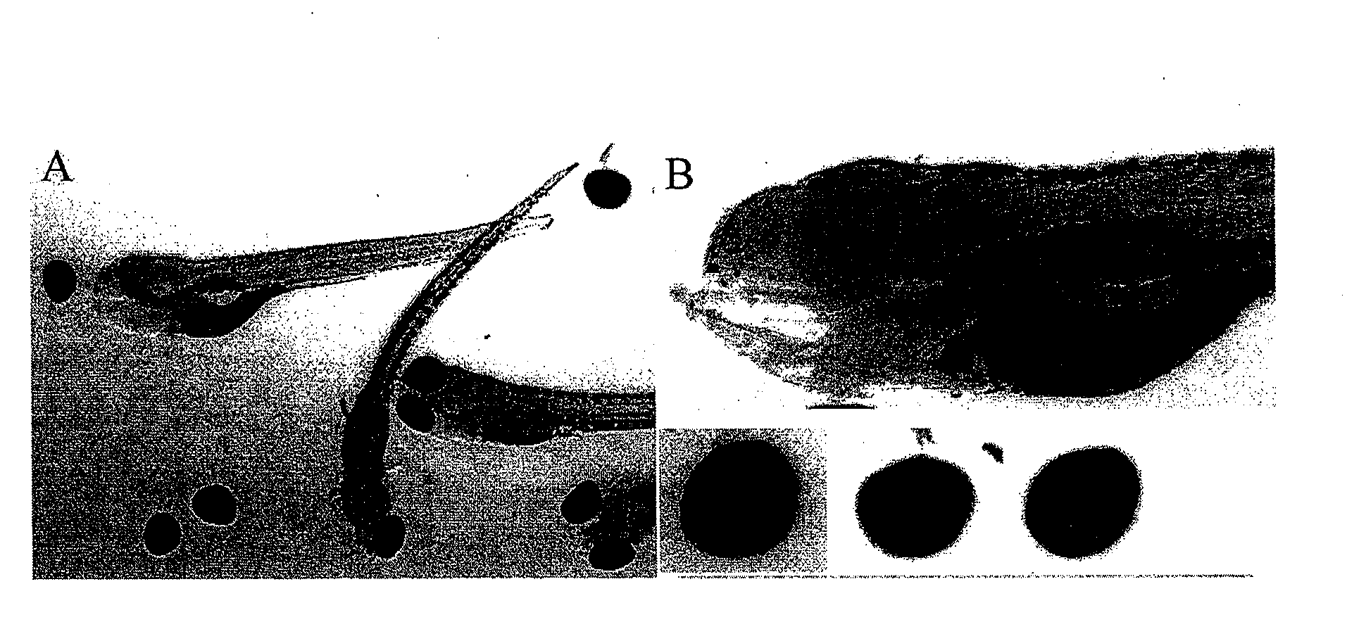
[0274]A method to rapidly dissociate eyes from whole mount immunostained zebrafish was developed by incubating zebrafish with a collagenase enzyme (Clostridium histolyticum collagenase Type II, Gibco) at various enzyme concentrations and temperatures, and for various periods of time. The enzyme concentrations ranged from 15 U / ml up to 150 U / ml. The temperature ranged from room temperature to 37° C. The period of incubation ranged from 30 minutes up to 16 hours. It was found that the enzyme concentration, temperature and period of time have to be coordinated to dissociate intact eyes. Using this collagenase, 150 U / ml of enzyme at 37° C. for 45 minutes and dissociating eyes (see FIG. 1) by gentle pipetting was effective in dissociating intact eyes. Using this approach, the vascular structure in the choroidal vessel plexus can be easily ...


PUM

PropertyMeasurementUnit
Structureaaaaaaaaaa
Sizeaaaaaaaaaa
Densityaaaaaaaaaa

Abstract

The present invention provides methods of screening an agent for an activity in an isolated organ, e.g., eye, from a teleost, e.g., zebrafish. Methods of isolating eyes from zebrafish are provided. Methods of screening an agent for an ocular activity in the isolated eye are provided. Methods of screening an agent for an ocular activity in a model of ocular disease or disorder are provided. Methods of screening an agent for an ocular activity in the isolated eye and for screening the agent for cell death and / or toxic activity in the eye or other organ or tissue are provided. The invention further provides high throughput methods of screening agents for an activity in isolated eyes of zebrafish in multi-well plates.

Description

CROSS-REFERENCE TO RELATED APPLICATIONS[0001]The present application is a nonprovisional of 60 / 934,455 filed Jun. 12, 2007, which is incorporated by reference in its entirety for all purposes.STATEMENT AS TO RIGHTS TO INVENTIONS MADE UNDER FEDERALLY SPONSORED RESEARCH AND DEVELOPMENT[0002]This invention was supported by grants from the National Institutes of Health (Grant Nos. 1R43EY016254-01, and 1R44EY015335-02). The U.S. Government may have certain rights in this invention.BACKGROUND OF THE INVENTION[0003]The identification of therapeutic or prophylactic compounds that may be useful for treating or preventing ocular diseases and disorders while exhibiting low toxicity and / or side effects is a focal point of drug discovery and development. Evaluation of the potential impact of drug candidates on human and animal health is a major component of the risk / benefit assessment. A number of cell-based in vitro efficacy and toxicity screens have been developed; these screens, however, do n...

Claims


Application Information

Patent Timeline
IPC IPC(8): A61K49/00
CPCA61K49/0008C12Q1/34G01N33/5088C12Q1/6876C12Q1/37
Inventor MCGRATH, PATRICIASENG, WEN LIN
Owner PHYLONIX PHARMA
Why Eureka
Try Eureka