


Quick Research
Generate reliable direction feasibility study reports for your R&D in just a few steps.

Technical Q&A
Discover and master advanced knowledge NOW. Basics, ideas, possibilities, all at once.

Find Solutions
As an expert in R&D theories, this can generate solutions to your technical problems instantly.

Evaluate Feasibility
Analyze your overall solution with one click, know your potential R&D risks in advance.

Monitor Landscape
Get weekly tech updates, stay abreast of the latest tech innovations and key insights.
Poly(ethylene glycol) Anti-body detection assays and kits for performing thereof
a technology of polyethylene glycol and antibody detection, which is applied in the field of polyethylene glycol antibody detection assays and kits for performing thereof, can solve the problems of ineffective peg-conjugated therapeutics, severe hypersensitivity reactions, and rapid clearance of substances from the body by the immune system,
- Summary
- Abstract
- Description
- Claims
- Application Information
AI Technical Summary
Problems solved by technology
Method used
Image

Examples
example 1
Testing For An Antibody To Poly(Ethylene Glycol):
Method:
Preparation of Antigen:
[0048]Blood type O red blood cells (RBCs) were washed 3 times with phosphate buffered saline (PBS, pH 7.4, 290 mOsm / kg) at 1400×g for 6 minutes. RBCs were then resuspended to a 10% hematocrit (hct) in 15 mM triethanolamine buffer (pH 8.4, 290 mOsm / kg). Poly(ethylene glycol) coating of RBCs was achieved by the addition of a reactive PEG to the RBC suspension. A succinimidyl propionate derivate of monomethoxy-poly(ethylene glycol) of molecular mass 20 kDa (mPEG20 k-SPA) was dissolved in cold 10 mM hydrochloric acid +154 mM NaCl, and added to the RBC suspension to yield a suspension phase concentration of 5 mg / mL mPEG20 k-SPA. The mixture was incubated at room temperature for 1 hour, and then washed 3 times with PBS at 500×g for 10 minutes. PEG-RBCs were then resuspended to a 5% hct in PBS and used for serologic testing.
Tube Test for Anti-PEG:
[0049]One drop of RBCs (PEG-coated or control (uncoated) RBCs) at ...
example 2
Gel Test For An Antibody To Poly(Ethylene Glycol)
Preparation of Antigen:
[0052]PEG-coated RBCs were prepared as described in Example 1.
[0053]Preparation of Gel Test tubes:
[0054]Gel Test tubes were prepared as follows. One hundred microliters of Sephaeryl 500-HR beads at 50% solids were pipetted into a narrow 300 μL tube. The tube was centrifuged at 500×g for 10 minutes.
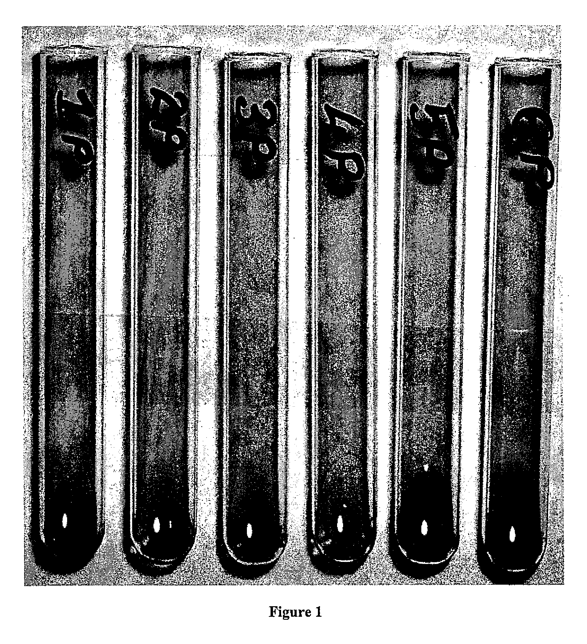
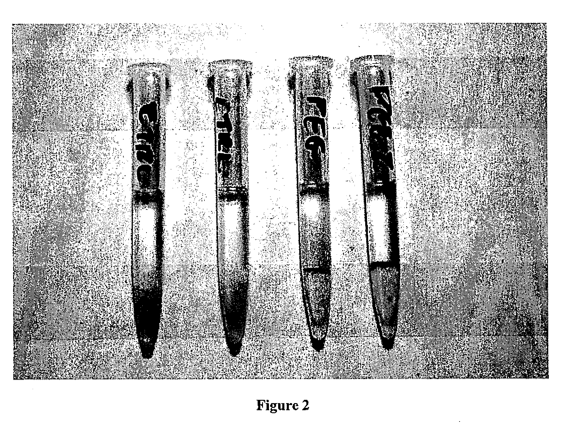
Example of Gel Test for Anti-PEG:
[0055]One hundred microliters of plasma was then pipetted on the top of the gel layer. Twenty five microliters of PEG-RBCs (or control (uncoated) RBCs) at 10% hct were added to the top of the plasma layer. Samples were incubated at room temperature for 15 minutes, and then centrifuged at 500×g for 3 minutes
[0056]Agglutination was scored accordingly: RBCs do not enter top of gel =4+ positive test, RBCs pass through gel=0 negative test.
Results:
[0057]The results of the gel test with PEG-RBCs are shown in FIG. 2. All uncoated (control) RBCs passed through the gel and gave a negative gel tes...
example 3
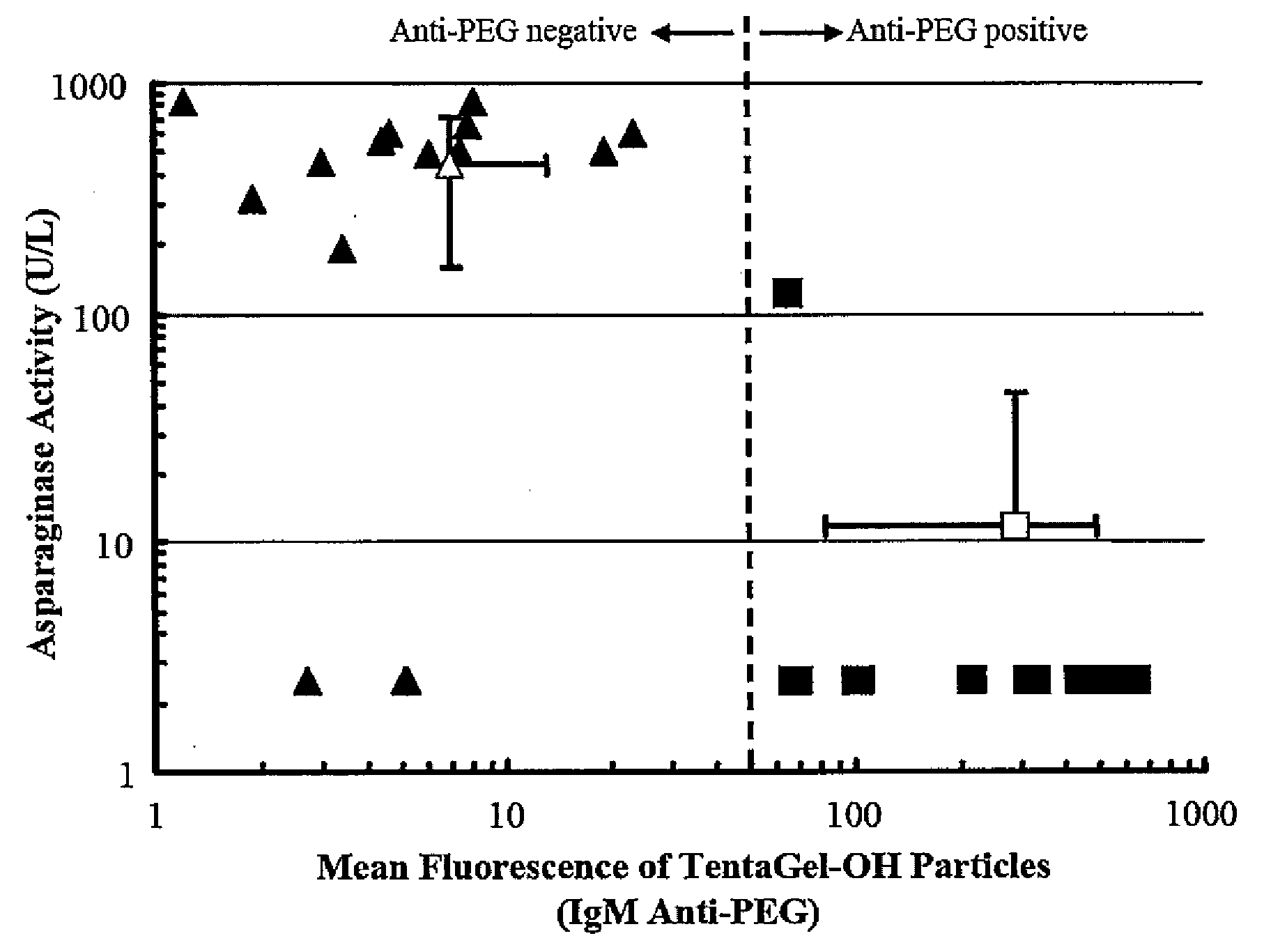
Flow Cytometric Testing For An Antibody To Poly(Ethylene Glycol) Method:
[0058]Fifty microliters of each test plasma were added to 100 μL of PBS and 25 μL of a 1% suspension of 10 μm diameter poly(ethylene glycol) particles, known commercially as TentaGel-OH beads (TentaGel-OH M 30 100, Rapp Polymere GmbH, Tübingen, Germany), which are composed primarily of PEG. The mixture was incubated for 1 hour at room temperature and the beads were washed twice with PBS (200×g for 2 minutes) and resuspended with 1 mL of PBS containing 5 μL of fluorescein isothiocyanate labeled-anti-human IgG and 5 μL of R-phycoerythrin labeled-anti-human IgM. After 1 hour incubation at room temperature in the dark, the particles were washed 3 times with PBS (200×g for 2 minutes) and resuspended with 0.5 mL of PBS and examined by flow cytometry. Ten thousand counts were recorded per sample, gated for single beads. Non-specific protein uptake was investigated by staining with FITC-anti-human albumin.
Results:
[0059]...
PUM
| Property | Measurement | Unit |
|---|---|---|
| diameter | aaaaa | aaaaa |
| diameter | aaaaa | aaaaa |
| diameter | aaaaa | aaaaa |
Abstract
Description
Claims
Application Information
 - R&D Engineer
- R&D Manager
- IP Professional
- Industry Leading Data Capabilities
- Powerful AI technology
- Patent DNA Extraction
Browse by: Latest US Patents, China's latest patents, Technical Efficacy Thesaurus, Application Domain, Technology Topic, Popular Technical Reports.
© 2024 PatSnap. All rights reserved.Legal|Privacy policy|Modern Slavery Act Transparency Statement|Sitemap|About US| Contact US: help@patsnap.com