
Rapid detection nanosensors for biological pathogens

a biological pathogen and nanosensor technology, applied in the field of rapid detection nanosensors for biological pathogens, can solve the problems of zero or near zero false positives, achieve high signal strength, increase sensitivity, and reduce background noise

Inactive Publication Date: 2010-04-29
EPIR TECH INC
View PDF1 Cites 14 Cited by
  • Summary
  • Abstract
  • Description
  • Claims
  • Application Information

AI Technical Summary

Benefits of technology

[0014]One advantage of the present invention is that it provides for the detection of pathogens in the order of minutes, rather than hours, and provides the ability to detect multiple pathogens simultaneously. Additionally, since QDs emit light with a higher intensity than fluorescent dyes, the invention gives high signal strength and near zero background noise which gives increased sensitivity over the prior art.

Problems solved by technology

When combined with the use of filters, the combination of quenchers and the QDs gives an increased signal to noise ratio resulting in zero or near zero false positives.

Method used


Image

  • Rapid detection nanosensors for biological pathogens
  • Rapid detection nanosensors for biological pathogens
  • Rapid detection nanosensors for biological pathogens

Examples


example 1

Antibody / QD Attachment

[0043]We first describe the preparation of the assay test solution used in the detection of the biological contaminants. For this study, E. coli 0157:H7 monoclonal antibodies (purchased from Biocompare Inc.) were conjugated to 605 nm QDs, B. cereus antibodies (purchased from Research Diagnostics Inc.) were conjugated to 565 nm QDs, and MS-2 virus antibodies (purchased from Tetracore Inc.) were conjugated to 525 nm QDs. All of the QDs used were CdSe / ZnS carboxyl coated Evitags™ (purchased from Evident Technologies, NY).

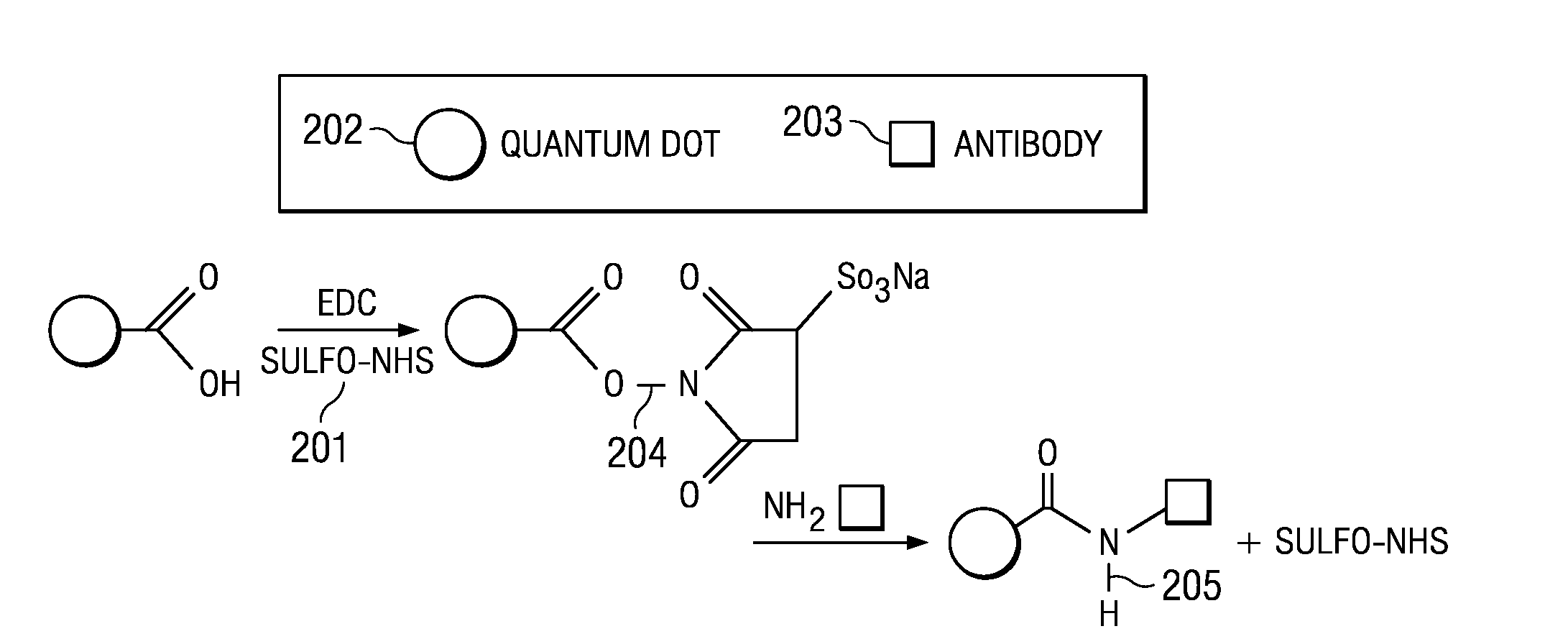
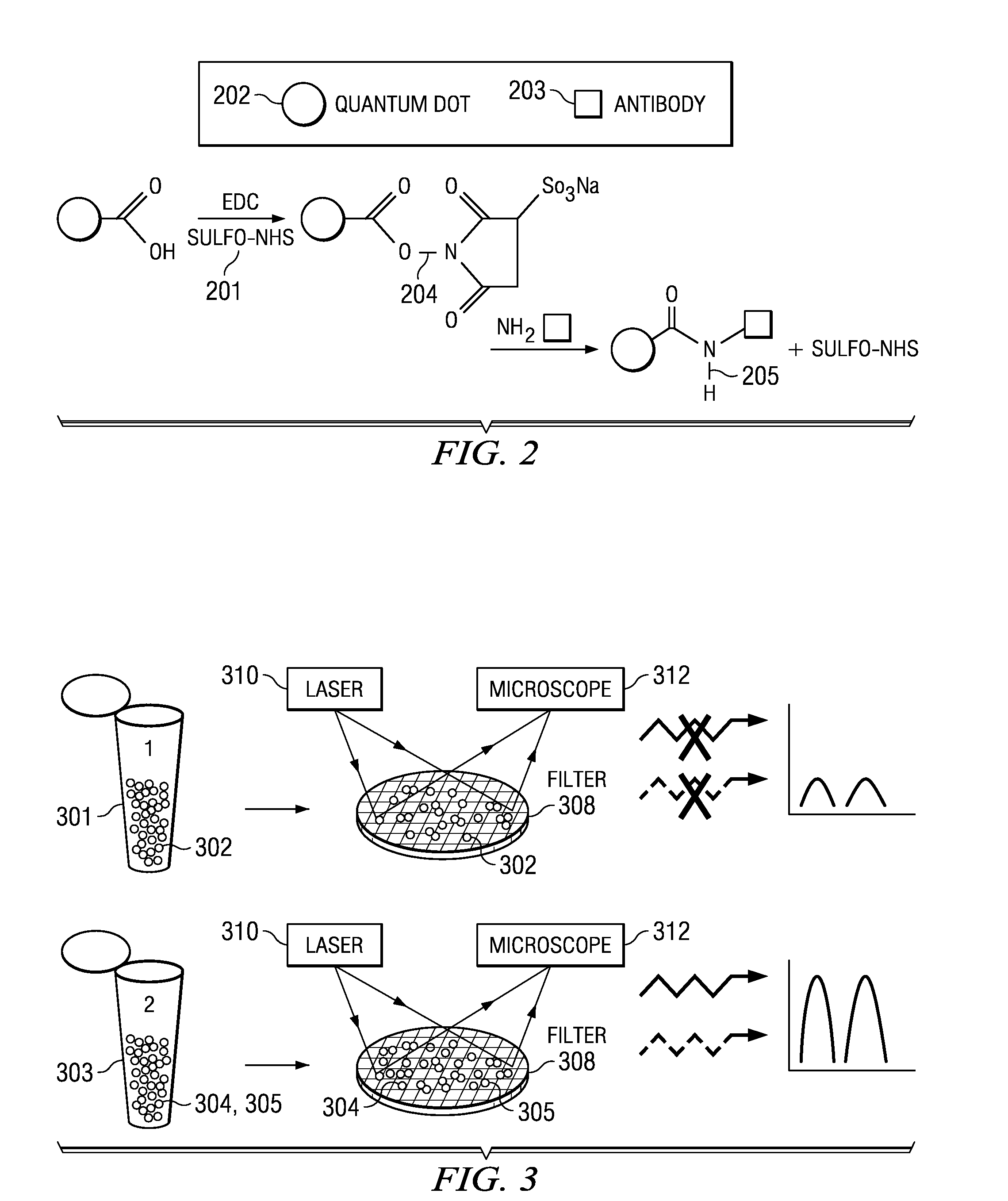
[0044]FIG. 2 shows the cross-linking reagents 201 used in functionalizing the QDs 202. Reagents 201 contain reactive ends for specific functional groups (amine and carboxyl groups). Cross-linking agents useful for binding biomolecules to QDs include EDC (1-ethyl-3-(3-dimethylaminopropyl) carbodiimide hydrochloride) and sulfo-NHS(N-hydroxysuccinimide). In the case of QDs functionalized with carboxyl groups, EDC reacts with the carboxylic acid group...

example 2

Inactivated Target Antigen / Quencher Attachment

[0046]Using similar procedures, carboxyl terminated quenchers were conjugated to inactivated E. coli 0157:H7 (purchased from KPL Inc. of Gaithersburg, Md.), inactivated B. cereus (purchased from ATCC Inc. of Manassas, Va.) and MS-2 antigens (obtained from CERL, Champaign, Ill.). Stock solutions were prepared containing 106 colony forming units per milliliter (CFUs / mL) of each type of antigen in standard buffers, and were stored separately according to the manufacturer's specifications before the conjugation reactions. The quencher chosen for the experiment was purchased from Biosearch Technologies of Novato Calif. Specifically the Black Hole Quencher-2 (BHQ-2). BHQ-2 was chosen for its strong absorbance (quenching) over a wide range of wavelengths in the visible region, making it suitable for multiplexing, in which QDs having different emission wavelengths are used for detection. Black Hole Quenchers are organic quencher compounds that o...


PUM


Abstract

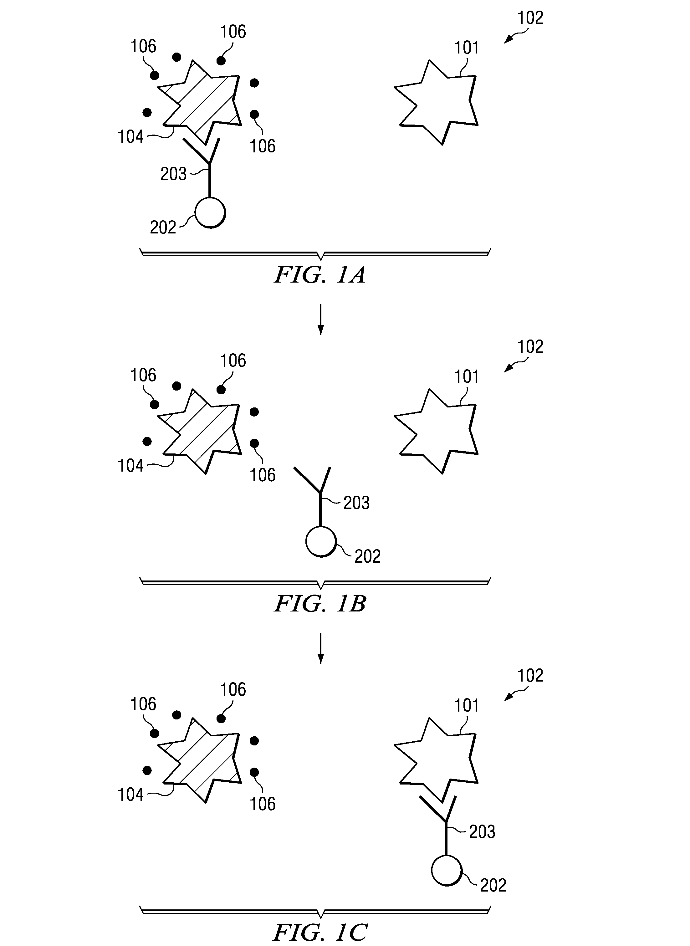
An assay test solution, a method for using, and an apparatus for the rapid detection of multiple pathogens using a FRET-based phenomenon. A volume of fluid, possibly containing pathogens, is passed through an intake and combined with an assay solution of quantum dot / antibody-antigen / quencher complexes that dissociate and recombine with the pathogens into quantum dot / antibody-pathogen complexes. The quantum dot / antibody-antigen / quencher and quantum dot / antibody-pathogen complexes are captured on a detection filter which is illuminated by a light source. The quantum dot / antibody-pathogen complexes, but not the quantum dot / antibody-antigen / quencher complexes, fluoresce when excited by the light from the light source and the fluorescence is picked up by a photodetector, indicating the presence of the pathogens.

Description

STATEMENT AS TO RIGHTS IN INVENTIONS MADE UNDER FEDERALLY-SPONSORED RESEARCH AND DEVELOPMENT[0001]This invention was made with Government support under Contract No. W9132T-06-C-0032 awarded by the United States Army. The government has certain rights in this invention.BACKGROUND OF THE INVENTION[0002]Political and social upheaval over the last decade combined with the dangers of chemical agents and biological pathogens have given the United States and other countries around the world an increasing cause for concern about the use of these agents against both citizens and military personnel. As a result, these concerns about public health and safety have fostered research into security systems that can detect these agents quickly and effectively.[0003]In addition to security applications, increasing limitations on the emissions of chemicals and biological agents to the environment require quick and accurate detection as a part of monitoring and compliance programs. Thus, there is a ne...

Claims


Application Information

Patent Timeline
IPC IPC(8): G01N33/569G01N33/53C12M1/00
CPCG01N33/569G01N33/542
Inventor RAMADURAI, DINAKARGARLAND, JAMES W.SIVANANTHAN, SIVALINGAM
Owner EPIR TECH INC
Learn More