
Bone Graft

a bone graft and bone graft technology, applied in the field of bone grafts, can solve the problems of demineralized bone matrix, limited abg amount, 5% surgical morbidity, etc., and achieve the effect of reducing activation of osteoinductive agents, reducing osteoinductive capacity, and improving shape-retaining characteristics

Active Publication Date: 2008-06-19
WARSAW ORTHOPEDIC INC
View PDF99 Cites 37 Cited by
  • Summary
  • Abstract
  • Description
  • Claims
  • Application Information

AI Technical Summary

Benefits of technology

The present invention provides improved demineralized bone matrix (DBM) compositions with better stability and osteoinductivity. The invention addresses the problem of fast reduction in osteoinductive capabilities of previous DBM compositions due to degradation of osteoinductive agents, diffusion of these agents out of the composition, and inhibition of their activation. The invention provides methods of preparing and using the improved DBM compositions, as well as systems and reagents for preparing and applying the compositions. The invention also provides a system for characterizing and identifying improved DBM compositions. Overall, the invention provides a more stable and effective DBM composition for bone regeneration and healing.

Problems solved by technology

Unfortunately, the amount of ABG available is limited, and there is at least a 5% surgical morbidity associated with the harvesting procedure.
One of the major disadvantages to demineralized bone matrix is that it often does not hold the three dimensional space of the defect.

Method used


Image

  • Bone Graft
  • Bone Graft
  • Bone Graft

Examples


example 1

Preparing Demineralized Bone Matrix (DBM)

[0092]DBM may be prepared using any method or technique known in the art (see Russell et al. Orthopedics 22(5):524-531, May 1999; incorporated herein by reference). The following is an exemplary procedure for preparing demineralized bone derived from Glowacki et al. “Demineralized Bone Implants” Clinics in Plastic Surgery 12(2):233-241, April 1985, which is incorporated herein by reference. Bones or bone fragments from donors are cleaned to remove any adherent periosteum, muscle, connective tissue, tendons, ligaments, and cartilage. Cancellous bone may be separated from dense cortical bone and processed as large pieces. Cortical bone may be cut into small pieces to improve the efficiency of subsequent washes and extractions. Denser bone from larger animals may need to be frozen and hammered in order to produce chips less than 1 cm. The resulting pieces of bone are thoroughly washed with cold, deionized water to remove marrow and soft tissue.

[...

example 2

Another Method of Preparing DBM

[0097]DBM may be prepared using any method or techniques known in the art (See Russell et al. Orthopedics 22(5):524-531, May 1999; incorporated herein by reference).

[0098]Demineralized bone matrix was prepared from long bones. The diaphyseal region was cleaned of any adhering soft tissue and then ground in a mill. Ground material was sieved to yield a powder with particles approximately 100 .mu.m to 500 .mu.m in diameter. The particulate bone was demineralized to less than about 1% (by weight) residual calcium using a solution of Triton X-100 (Sigma Chemical Company, St Louis, Mo.) and 0.6N HCl at room temperature followed by a solution of fresh 0.6N HCl. The powder material was rinsed with deionized water until the pH was greater than 4.0. It then was soaked in 70% ethanol and freeze-dried to less than 5% residual moisture.

example 3

Formulating Preferred Inventive DBM Compositions

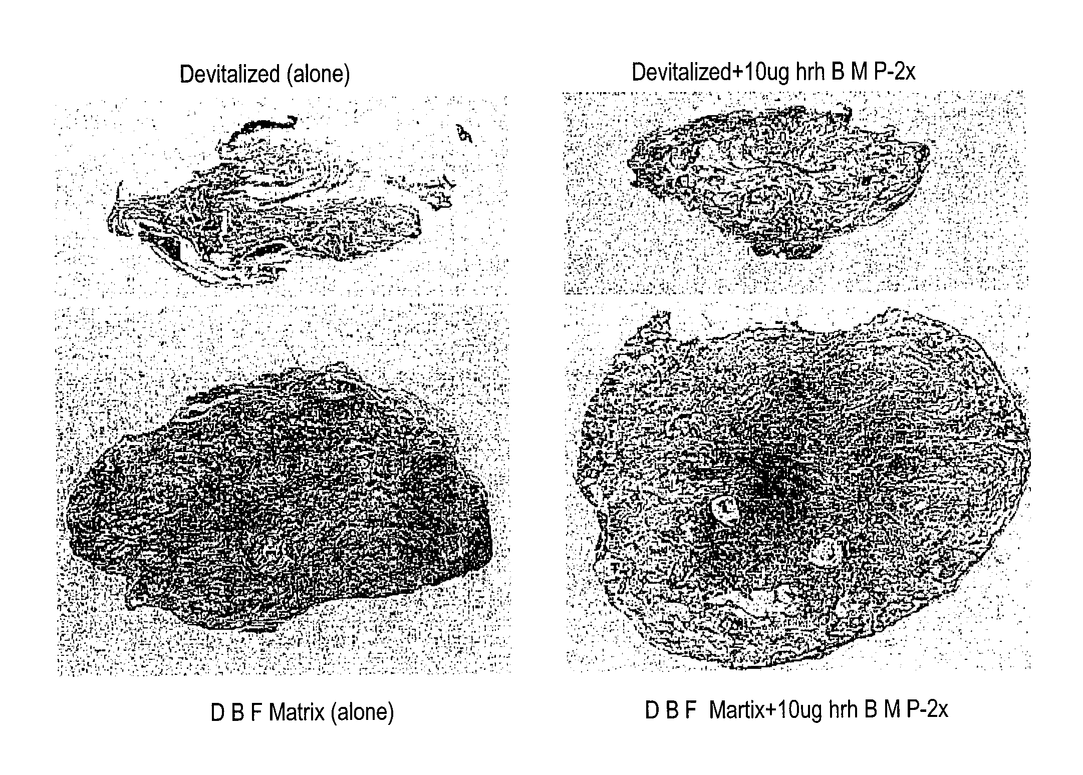
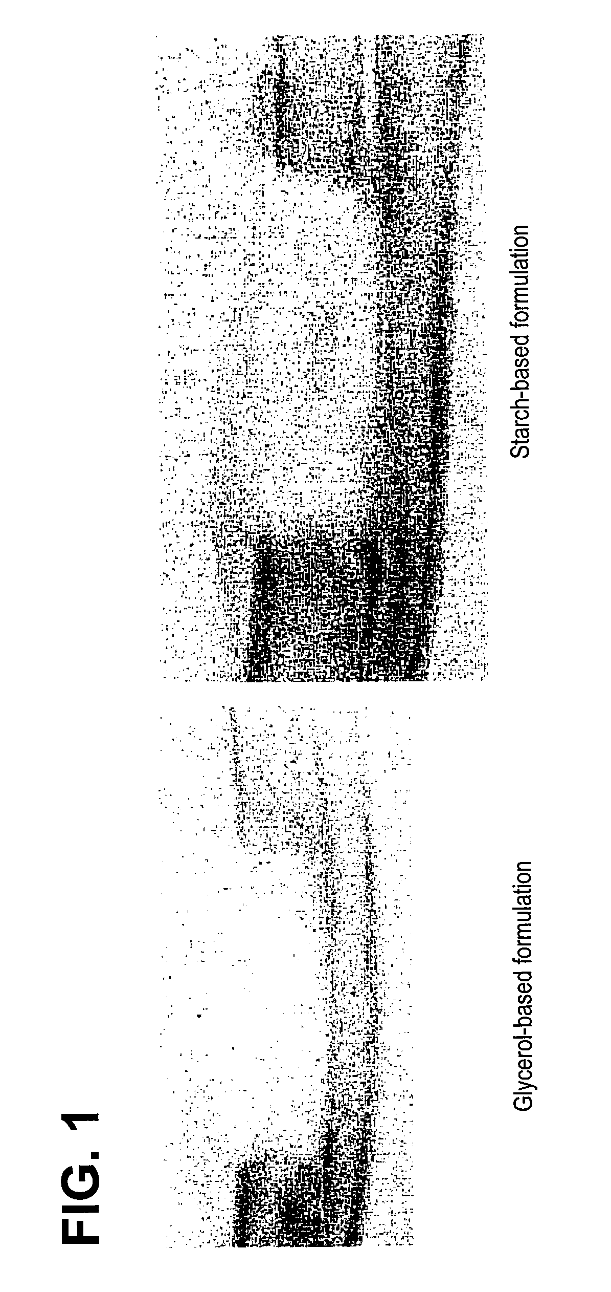
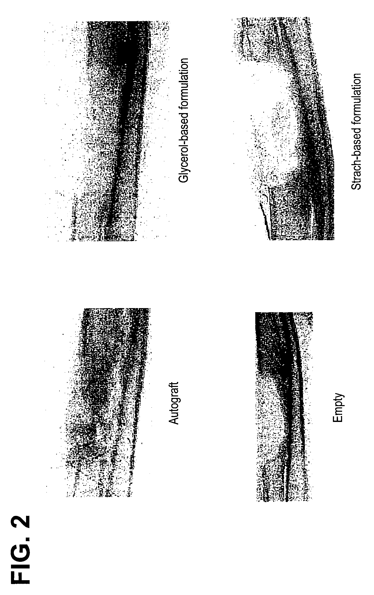
[0099]The carrier was prepared by mixing approximately 6.5% (w / w) of the modified starch, B980, with approximately 30% (w / w) maltodextrin (M180) and approximately 63.5% (w / w) sterile, deionized water. The mixture was heated to 70 .degree. C. to pre-gelatinize. The pre-gelatinized mixture was then transferred into a steam autoclave and sterilized / gelatinized at 124 .degree. C. for 2 hours. The resulting mixture then had a consistency of pudding. The cooled carrier mixture was then combined with DBM (from Example 2) and water, in a ratio of approximately 27:14:9, respectively. The stabilized DBM was then implanted into athymic rats to assess osteoinductivity.

[0100]Alternative embodiments: Other components such as glycerol were added as a solution (approximately 20% w / v) in water instead of water at the time of pre-gelatinization or during the final composition mixing and were found to have acceptable handling characteristics.


PUM

PropertyMeasurementUnit
sizeaaaaaaaaaa
sizeaaaaaaaaaa
sizeaaaaaaaaaa

Abstract

An improved demineralized bone matrix (DBM) or other matrix composition is provided that has been mixed with a stabilizing agent that acts as (1) a diffusion barrier, (2) a enzyme inhibitor, (3) a competitive substrate, or (4) a masking moiety. A diffusion barrier acts as a barrier so as to protect the osteoinductive factors found in DBM from being degraded by proteolytic and glycolytic enzymes at the implantation site. Stabilizing agents may be any biodegradable material such as starches, modified starches, cellulose, dextran, polymers, proteins, and collagen. As the stabilizing agents degrades or dissolves in vivo, the osteoinductive factors such as TGF-.beta., BMP, and IGF are activated or exposed, and the activated factors work to recruit cells from the preivascular space to the site of injury and to cause differentiation into bone-forming cells. The invention also provides methods of preparing, testing, and using the inventive improved osteodinductive matrix compositions

Description

CROSS-REFERENCE TO RELATED APPLICATIONS[0001]This is a divisional application of U.S. patent application Ser. No. 10 / 271,140, filed on Oct. 15, 2002, which claims priority to U.S. Application Ser. No. 60 / 392,462, filed Jun. 27, 2002, and U.S. Application Ser. No. 60 / 329,156, filed Oct. 12, 2001, the contents of which are incorporated herein by reference in its entirety.BACKGROUND[0002]The rapid and effective repair of bone defects caused by injury, disease, wounds, surgery, etc., has long been a goal of orthopaedic surgery. Toward this end, a number of compositions and materials have been used or proposed for use in the repair of bone defects. The biological, physical, and mechanical properties of the compositions and materials are among the major factors influencing their suitability and performance in various orthopaedic applications.[0003]Autologous cancellous bone (“ACB”) is considered the gold standard for bone grafts. ACB is osteoinductive, is non-immunogenic and, by definitio...

Claims


Application Information

Patent Timeline
Patent Type & Authority Applications(United States)
IPC IPC(8): A61F2/28A61K35/32A61P19/00A61F2/30A61M31/00A61L27/00A61L27/22A61L27/36A61L27/50A61L27/54A61L27/58
CPCA61F2002/2817A61L2430/02A61F2002/30677A61L27/227A61L27/3608A61L27/3633A61L27/365A61L27/505A61L27/54A61L27/58A61L2300/414A61L2300/434A61L2300/602A61L2300/802A61F2002/2835B33Y80/00A61P19/00A61L27/44B82Y5/00A61F2/28
Inventor KNAACK, DAVIDTRAIANEDES, KATHYDIEGMAN, MICHELEFORSYTH, NANETTEWINTERBOTTOM, JOHN
Owner WARSAW ORTHOPEDIC INC
Learn More