
Animal models of tumor metastasis and toxicity

Inactive Publication Date: 2007-12-06
PROJECH SCI TO TECH
View PDF0 Cites 19 Cited by
  • Summary
  • Abstract
  • Description
  • Claims
  • Application Information

AI Technical Summary

Benefits of technology

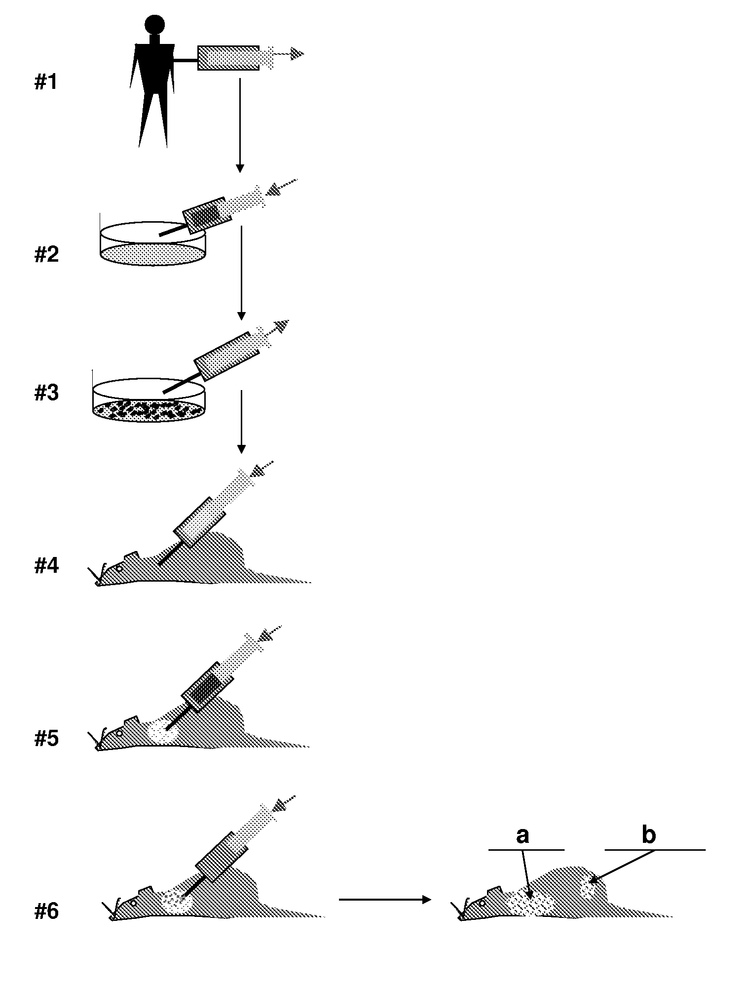
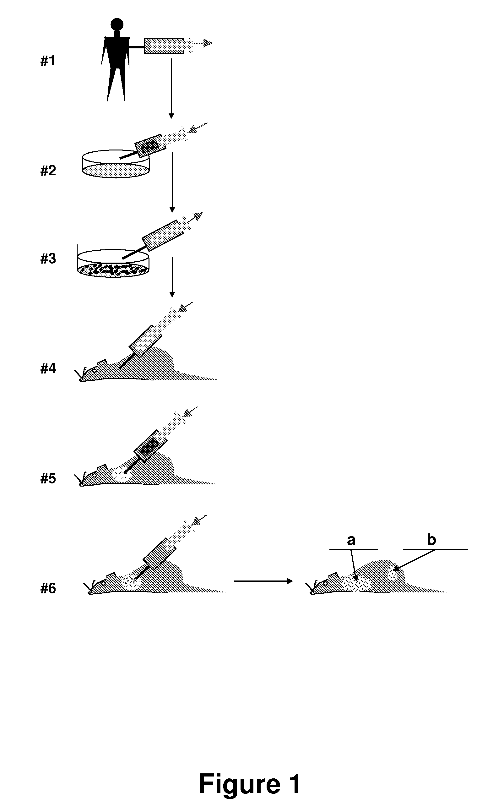
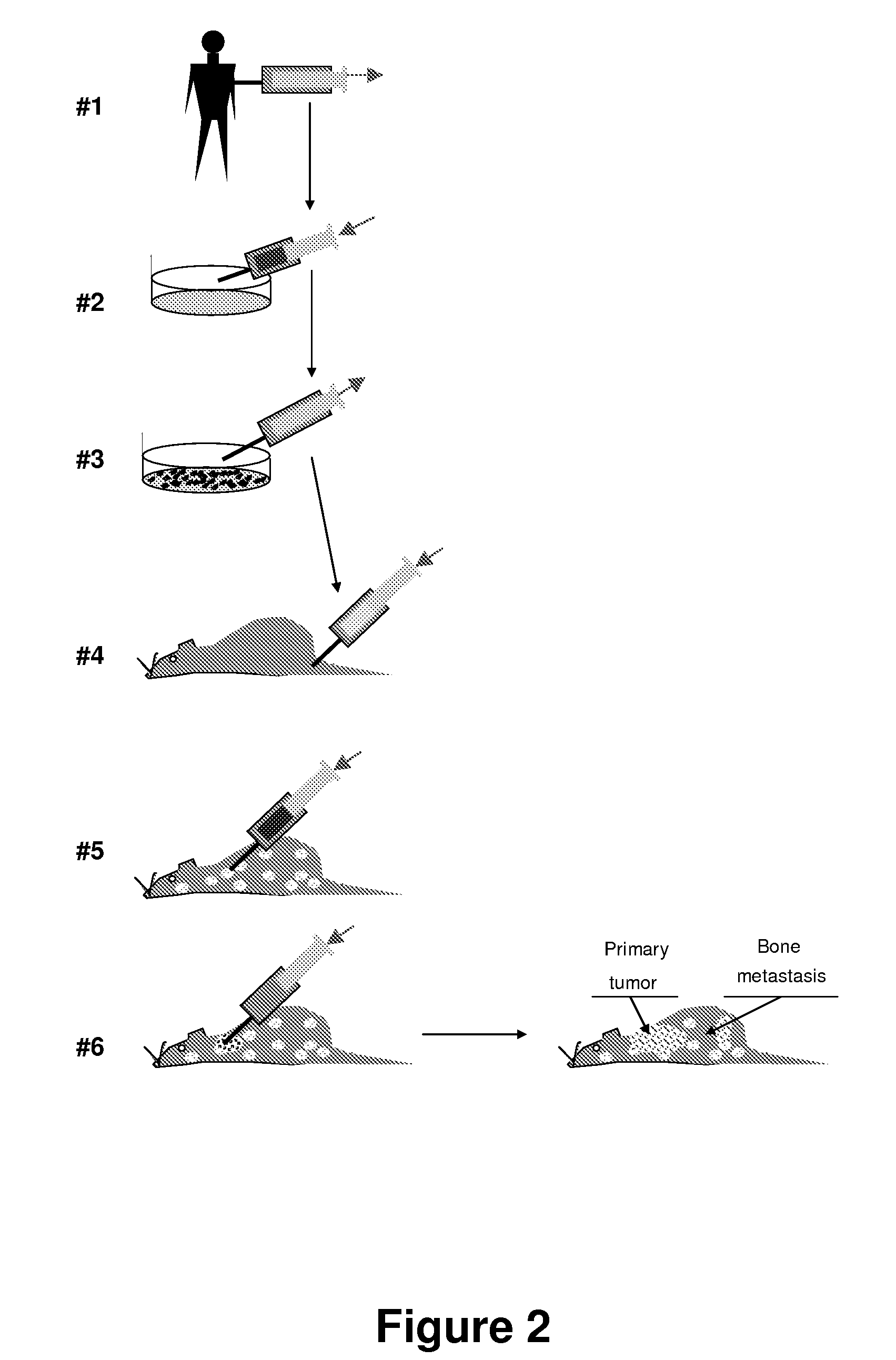
[0015]The present invention is based on the surprising finding that the targeting efficiency of the stem cells into an organ of choice can be substantially improved if the recipient animal, prior to receiving the tumor cells, has been treated so as to inflict damage leading to cellular death to the tissue in the recipient organism which corresponds to the target tissue is damaged. In this manner, the damaged organ can be sensed by the implanted stem cells that migrate to the site of damage, undergo differentiation, and promote structural and functional repair or that organ. The animals carrying said reconstituted organ are exceptionally useful as experimental models to study metastasis processes as well as for studying interactions between exogenous compounds and said organs.

Problems solved by technology

However, the use of these models for studying the metastases of human cancer cells has so far been limited, due to (i) the low efficiency of the incidence of cancer metastasis in the recipient mice, usually due to the fact that the transplanted material consists of tumor derived cell lines that often are cells that have been over transformed in vitro after many uncontrolled passages and (ii) the large cell number required to achieve the desired results.
These cells (also called cancer stem cells) have been well characterized in human breast cancer but are difficult to isolate.
However, this type of models poses different challenges which make them not easy to implement.
Although it has been demonstrated that human stem cells injected into the mouse blood stream, can localize to multiple organs and have the capacity to recapitulate the local cellularity of each organ, these methods are faced with the problem that the transplanted stem cells, depending on the injection location or method, localize to a variety of organs, so that it is often difficult reconstitute in the recipient animal the organ of interest.

Method used


Image

  • Animal models of tumor metastasis and toxicity
  • Animal models of tumor metastasis and toxicity
  • Animal models of tumor metastasis and toxicity

Examples


example 1

Isolation of Tissues from Human Fetal Tissues and Grafting into Immunodeficient Animals (SCID-hu Mice).

[0129]C.B-17 scid / scid mice are bred, treated with antibiotics, as is well known in the art, and used at an age 6 to 8 weeks. Anaesthesia is used during all operative procedures. The human fetal tissues are derived from curettage operation involving physical extraction without administration of prostaglandins or related drugs. The tissues are individually placed in sterile 50 ml tubes containing RPMI 1640 medium supplemented with 10% fetal bovine serum (FBS), 50 U / mL penicillin, and 50 pg / mL streptomycin at 4° C. The samples are then shipped on wet ice, received within 16 to 20 hours, and transplanted into SCID mice within 36 hours. Cells from fetal thymus or liver are tested for the presence of HIV by the DNA polymerase chain reaction as described in all cases before use. Human fetal femurs and tibias are obtained at 17 to 22 gestational weeks (gw), when intramedullary hematopoies...

example 2

In Vivo Metastasis Assays in SCID-hu Mice

[0131]SCLC cells grown in vitro as suspension cultures were harvested by centrifugation, resuspended in Hanks' balanced saline solution (HBSS), assessed for cell number and viability, and injected into SCID-hu mice via lateral tail vein (experimental metastasis assay). For spontaneous metastasis assay cells were injected directly into one of the HFL grafts through a small incision in the skin.

[0132]Histology. Fragments of human grafts, murine internal organs (lungs, liver, spleen, adrenals, and sometimes additional organs), backbones, and sternums were dissected and fixed in buffered 20% (vol / vol) formalin. Bone tissues were treated with decalcifying solution (Baxter Scientific Products, McGaw Park, Ill.). After paraffin embedding, 4 □m sections are cut and stained with hematoxylin / eosin.

example 3

Engraftment of MSC Cells into a Recipient Animal

[0133]There is a rational to this technique: the expected multiorgan tropism of human mesenchymal stem cells in a variety of epithelia. Seeding of human MSC on different mouse organs can be confirmed using a variety of techniques well known in the art (immunostaining, human or mouse specific PCRs, etc.). In a variation of the strochimeric technique, around the time of the MSC injection (preferentially, before), mice can be treated with physical (gamma radiation) or chemical agents with cytostatic activity, in order to suppress the mobilization and proliferation of autologous BM MSC. This treatment could enrich the engrafted human MSC population in different organs. The treatment, in addition to down-regulate the normal proliferative response of autologous BM MSC to injury, can induce a generalized injury to dividing epithelial tissues. The latter effect is likely to serve as a stimulus to homing for injected human MSC


PUM


Abstract

A method for conversion of an animal into an appropriate recipient of tumor cells derived from a different species. Animals for such purpose can be immuno-incompetent animals that are doubly grafted with orthotopic tissues, in which one grafted tissue (i.e., breast) is from an organ of the same class as the tumor of origin (graft A), and the second grafted tissue (i.e., bone) is from a organ of the same class as a target organ for metastasis (graft B). These dual grafted animals can be used to model human diseases. In one implementation, human tumor cells are orthotopically seeded in graft A in order to analyze the occurrence of metastasis in graft B. Methods and compositions are described for creating a multiorgan human environment in mice, by grafting human stem cells (mesenchymal, embryonic or others) into mice, e.g., injured to enhance specific tissue engraftment. Such chimeric mice can be used to grow human tumors and to study the occurrence of metastasis.

Description

CROSS-REFERENCE TO RELATED APPLICATION[0001]This claims the benefit of priority under 35 USC §119 of U.S. Provisional Patent Application No. 60 / 809,306 filed May 31, 2006. The entire disclosure of said U.S. Provisional Patent Application No. 60 / 809,306 is hereby incorporated herein by reference in its entirety for all purposes.FIELD OF THE INVENTION[0002]The invention relates to the field of animal metastasis models and, in particular, to an animal metastasis model wherein the organ which is the target for metastasis is from a different species than the animal and has been reconstituted by damaging the corresponding organ in the animal prior to the administration of stem cells. Moreover, the invention relates also to the field of animals carrying xenoorgans and the uses thereof in toxicity models.BACKGROUND OF THE INVENTION[0003]Metastasis (Greek: change of the state, plural: metastases), sometimes abbreviated Mets, is the spread of cancer from its primary site (source tissue) to ot...

Claims


Application Information

Patent Timeline
IPC IPC(8): A01K67/027
CPCA01K67/0271A01K2227/105G01N2800/52G01N33/5088A01K2267/0331
Inventor RODRIGUEZ CIMADEVILLA, JUAN CARLOSPUNZON GAU, ISABEL
Owner PROJECH SCI TO TECH
Learn More