Method for predicting liver fibrosis and related pathologies
a liver fibrosis and pathology technology, applied in the field of medical diagnostics, can solve the problems of liver fibrosis development, high cost of liver biopsy, and associated costs, and achieve the effect of liver scarring
- Summary
- Abstract
- Description
- Claims
- Application Information
AI Technical Summary
Benefits of technology
Problems solved by technology
Method used
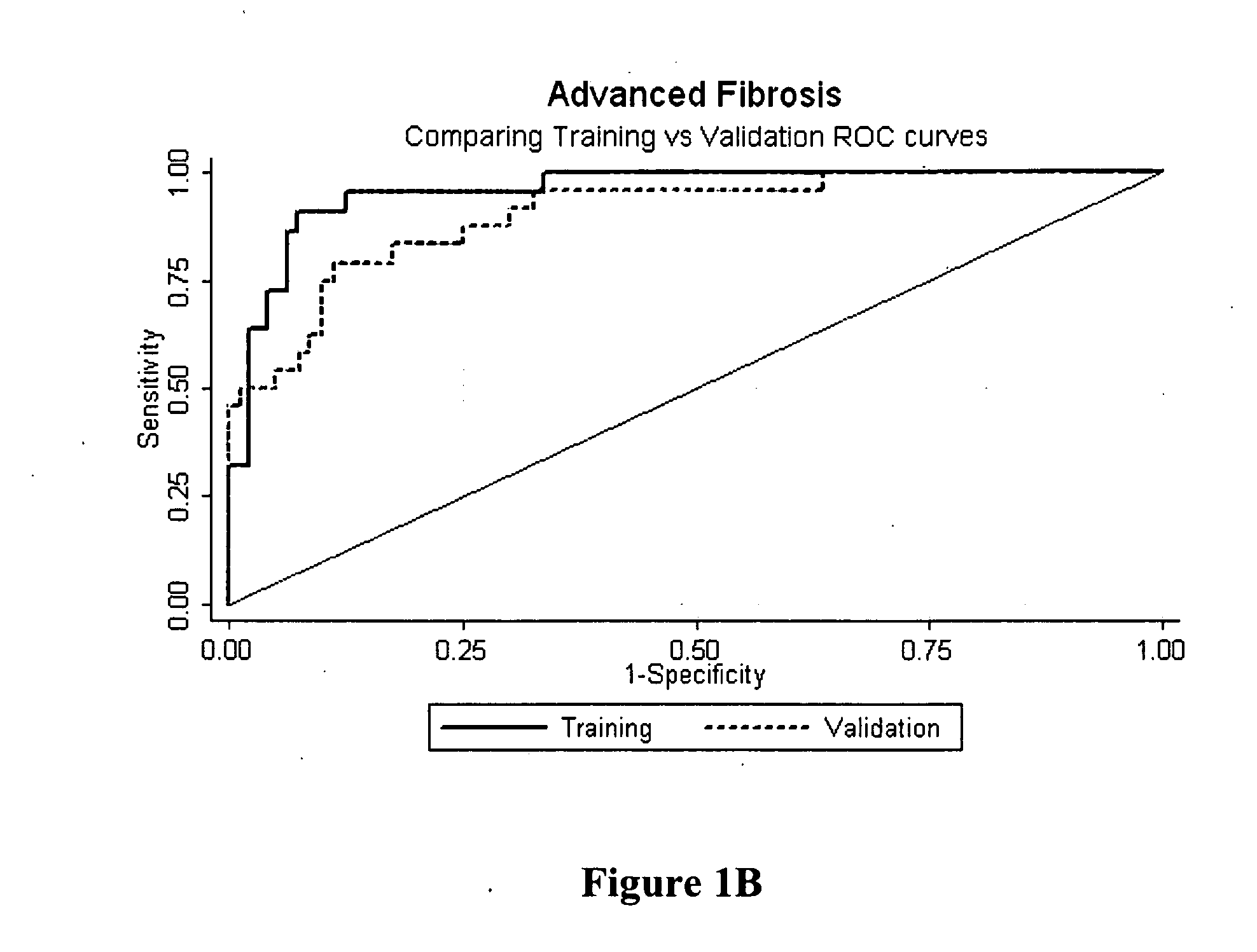
Image


Examples
example 1
[0091] Selection of Patient Population. Patients were prospectively recruited from viral liver clinics in different tertiary referral centers; the training set was recruited from Sir Charles Gairdner Hospital (Perth, Australia) and the validation set from Westmead Hospital and Royal Prince Alfred Hospital (Sydney, Australia). All patients had detectable hepatitis C RNA at the time of evaluation and were treatment naive. Coexisting liver disease due to hepatitis B, haemochromatosis, alpha-1 antitrypsin deficiency, Wilson's disease, autoimmune and cholestatic liver diseases were excluded by standard clinical, laboratory, imaging and histological studies. No patient had human immunodeficiency virus co-infection or had undergone liver transplantation. Liver biopsy was performed as part of the routine clinical care of these patients. Age, gender and viral genotype were recorded at time of liver biopsy. Patients from the training set had the Fibrotest calculated.
[0092] The demographic an...
example 2
[0093] Assay of Markers. Training set sera were analyzed for 10 candidate markers. Bilirubin, ALT, GGT, and albumin were all measured on fresh serum within 36 hours of collection using an automated biochemistry analyzer (Hitachi 917, Roche Diagnostics, Mannheim, Germany). Other analyses were performed in batches using frozen serum stored at −20 C. TIMP-1 and MMP-2 were measured by enzyme-linked immunosorbent assay on a 96-well microplate (Biotrak, Amersham Biosciences, Bucks, UK). Hyaluronic acid was measured by an enzyme-linked protein binding assay, also on a 96-well microplate (Corgenix, Westminster, Colo., US). Alpha-2 macroglobulin, apolipoprotein-A1 and haptoglobin were all obtained by nephelometry (Image, Beckman Coulter, Brea, Calif., US). All analyses were performed at a central laboratory, PathCentre in Perth.
[0094] Univariate logistic regression analysis of the variables tested in the training set revealed age, gender, albumin, hyaluronic acid, α-2 macroglobulin and TIMP...
example 3
[0096] Analysis of Liver Biopsy. Liver biopsies of both training and validation sets were a minimum of 18 gauge with a minimum of five portal tracts and were routinely stained with hematoxylin-eosin and trichrome stains. Biopsies were interpreted according to the scoring schema developed by the METAVIR group by two expert liver pathologists who were masked to patient clinical characteristics and serum measurements. Thirty biopsies were scored by both pathologists and interobserver agreement was calculated using Kappa (κ) statistics. Fibrosis was scored on the 5-point METAVIR scale. Necro-inflammatory activity, based on assessment of piecemeal and lobular necrosis, was graded on a 4-point scale as follows: AO, no activity; A1, mild; A2, moderate; A3, severe.
PUM
 Abstract
Description
Claims
Application Information
 - R&D
- Intellectual Property
- Life Sciences
- Materials
- Tech Scout
- Unparalleled Data Quality
- Higher Quality Content
- 60% Fewer Hallucinations
Browse by: Latest US Patents, China's latest patents, Technical Efficacy Thesaurus, Application Domain, Technology Topic, Popular Technical Reports.
© 2025 PatSnap. All rights reserved.Legal|Privacy policy|Modern Slavery Act Transparency Statement|Sitemap|About US| Contact US: help@patsnap.com