
Method and device for abrading skin

a technology of skin absorption and skin absorption, which is applied in the field of skin absorption methods and devices, can solve the problems of excessively reducing the skin barrier function, increasing the risk of infection, and pain, and achieves the effects of minimal irritation, simple and reliable manner, and efficient penetration of stratum corneum

Inactive Publication Date: 2006-11-23
BECTON DICKINSON & CO
View PDF4 Cites 67 Cited by
  • Summary
  • Abstract
  • Description
  • Claims
  • Application Information

AI Technical Summary

Benefits of technology

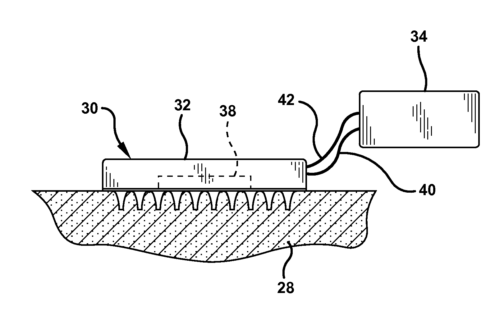
[0012] The present invention is directed to a method and device for abrading the skin, and particularly, the stratum corneum of the skin. The invention is further directed to a method of obtaining a sample or for the transdermal delivery of a substance, such as a drug or pharmaceutical agent, through the abraded area on the stratum corneum. One aspect of the invention is directed to a method and device for preparing a delivery site on the skin to enhance the delivery of a pharmaceutical agent through the stratum corneum of the skin to a sufficient depth where the pharmaceutical agent can be absorbed and utilized by the body.
[0014] Accordingly, a primary object of the invention is to provide a method and device for efficiently penetrating the stratum corneum substantially without pain to the patient and with a minimum of irritation to skin, thereby exposing the tissue below the stratum corneum directly to a pharmaceutical agent for absorption by the body.
[0015] A further object of the invention is to provide a method for abrading the stratum corneum in a simple and reliable manner.
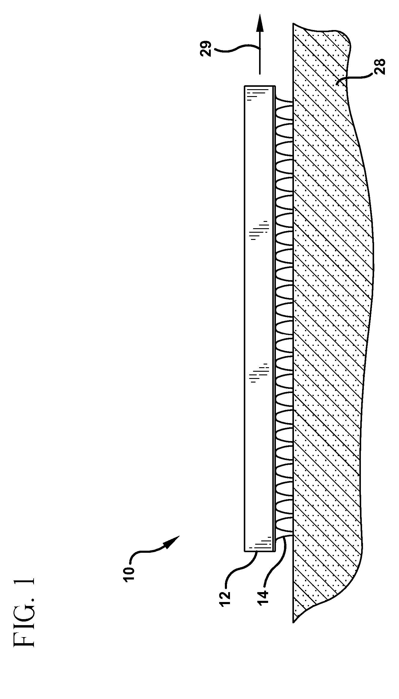
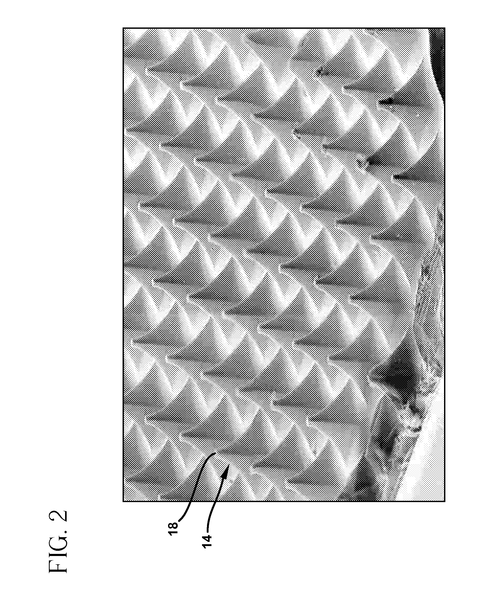
[0025] These and other objects of the invention are substantially achieved by providing a device for abrading the skin to promote the delivery or withdrawal of a substance through the skin of a patient. In a preferred embodiment, the device comprises a planar support having a bottom surface. A plurality of microneedles is coupled to and integral with the bottom surface of the support. The microneedles have a blunt, flat tip and a length sufficient to penetrate the stratum corneum of the skin without piercing the stratum corneum during abrading of the skin to enhance the permeability of the skin.

Problems solved by technology

These methods are usually painful or uncomfortable and increase the risk of infection by excessively reducing the skin barrier function.
These methods require complex and energy intensive electronic devices that are relatively expensive.
The chemical enhancers are often not suitable for transdermal drug delivery or sampling.
The amount and rate of drug delivery using iontophoresis can be difficult to control.
Although numerous efforts to enhance drug delivery using sonic energy have been proposed, the results generally show a low rate of drug delivery.
The prior methods and apparatus for the transdermal administration of drugs have exhibited limited success.

Method used


Image

  • Method and device for abrading skin
  • Method and device for abrading skin
  • Method and device for abrading skin

Examples


example 1

[0077] A microabrader having a surface area of about 1 cm2 is provided with a plurality of microneedles having a length of about 250 microns. The microneedles were arranged in a plurality of uniform rows and columns to provide a needle density of about 200 needles per cm2.

[0078] The microabrader was gently placed on the back of guinea pigs and moved across the skin to produce an abraded area of about 4 cm2. The microabrader was scraped along the same path several times to produce an abraded delivery site. The microabrader was removed and a commercially available anesthetic cream sold under the trademark EMLA was applied. The anesthetic cream was applied to a second group of guinea pigs in the same location without abrading.

[0079] The topical anesthetic was allowed to contact the skin for one hour before conducting the test for anesthesia. Each guinea pig received five controlled stimuli on the treatment site. In the negative control group, the test site was defined by a similar ci...

example 2

[0082] A microabrader having microneedles of about 200 microns in length was used to abrade the skin of guinea pigs in preparation for delivery of the anesthetic lidocaine by iontophoresis.

[0083] Iontophoresis patches were applied to the abraded delivery site to deliver lidocaine for 5 minutes at 1.8 mA. The control delivery sites without abrasion were treated with an identical lidocaine iontophoresis device for 5 minutes. The anesthesia obtained by the twitch method of Example 1 is presented in the graph of FIG. 8. The iontophoresis current was discontinued after 5 minutes and the extent of anesthesia measured for 1 hour. As shown by the data of FIG. 8, iontophoresis applied to a microabrasion site attained 100% anesthesia immediately after application, while the same iontophoresis without abrading attained about 50% anesthesia.

[0084] As shown in the graph of FIG. 8, the abraded site maintained a higher percent anesthesia than the site without abrasion.

example 3

[0085] This example evaluates the dose of lidocaine in the tissue. Lidocaine iontophoresis was conducted on anesthetized Yorkshire pigs using patches spiked with 14C lidocaine. Four abraders were selected having different microneedle lengths and shapes as follows: 100 microns with sharp points; 100 microns with blunt, flat tips; 200 microns with sharp points; and 200 microns with blunt, flat tips.

[0086] A delivery site was prepared on the pigs by abrading the skin with each of the microabraders and the patches were applied at about 1.8 mA. The radiolabeled lidocaine that was delivered to the pig was imaged on tape strips and assayed in the skin underlying the patch application site. The tape strips qualitatively show enhancement of lidocaine delivery with abrasion.

[0087] The treated skin was biopsied and cut into sections that were then dissolved and assayed for radiolabeled lidocaine with liquid scintillation counting. The average doses were determined by averaging the tissue dos...


PUM


Abstract

A device includes a plurality of microneedles for abrading the stratum corneum of the skin to form a plurality of grooves in the tissue having a controlled depth and width. The microneedles have a length of about 5-250 microns and generally about 5-200 microns. The device is rubbed over the skin to prepare an abraded site after which a transdermal delivery or sampling device is applied to the abraded delivery site. The abrasion increases the permeability of the skin and the rate of delivery and extraction of a substance without pain or irritation to the patient.

Description

[0001] This application is continuation of U.S. application Ser. No. 10 / 446,545, filed May 28, 2003, which is a divisional of U.S. application Ser. No. 09 / 405,488, filed Sep. 24, 1999.FIELD OF THE INVENTION [0002] The present invention relates to a method and device for abrading the skin. More particularly, the invention is directed to a method of abrading the stratum corneum to promote transdermal delivery or sampling of a substance. BACKGROUND OF THE INVENTION [0003] The skin is made up of several layers with the upper composite layer being the epithelial layer. The outermost layer of the skin is the stratum corneum that has well known barrier properties to prevent external molecules and various substances from entering the body and internal substances from exiting the body. The stratum corneum is a complex structure of compacted keratinized cell remnants having a thickness of about 10-30 microns. The stratum corneum forms a hydrophobic membrane to protect the body from invasion b...

Claims


Application Information

Patent Timeline
Patent Type & Authority Applications(United States)
IPC IPC(8): A61M31/00A61M37/00A61N1/30
CPCA61B17/205A61B17/54A61B2017/00761A61B2017/320004A61N1/303A61M2037/0007A61M2037/0046A61M2037/0053A61M2037/0061A61M37/0015
Inventor SAGE, JR., BURTON H.BOCK, CARL RANDOLPH
Owner BECTON DICKINSON & CO
Learn More