Methods of detecting breast cancer, brain cancer, and pancreatic cancer
a breast cancer and brain cancer technology, applied in the field of breast cancer, brain cancer and pancreatic cancer detection, can solve the problems of selective entrapment, inability to afford conventional imaging techniques such as computerized tomography and magnetic resonance imaging (mri), and inability to conclude the diagnosis of suspected breast cancer
- Summary
- Abstract
- Description
- Claims
- Application Information
AI Technical Summary
Benefits of technology
Problems solved by technology
Method used
Image
Examples
example 1
Specificity of NM404 for Neoplasia versus Hyperplasia in the ApcMin / + Endogenous Mammary Adenocarcinoma Model
Materials and Methods
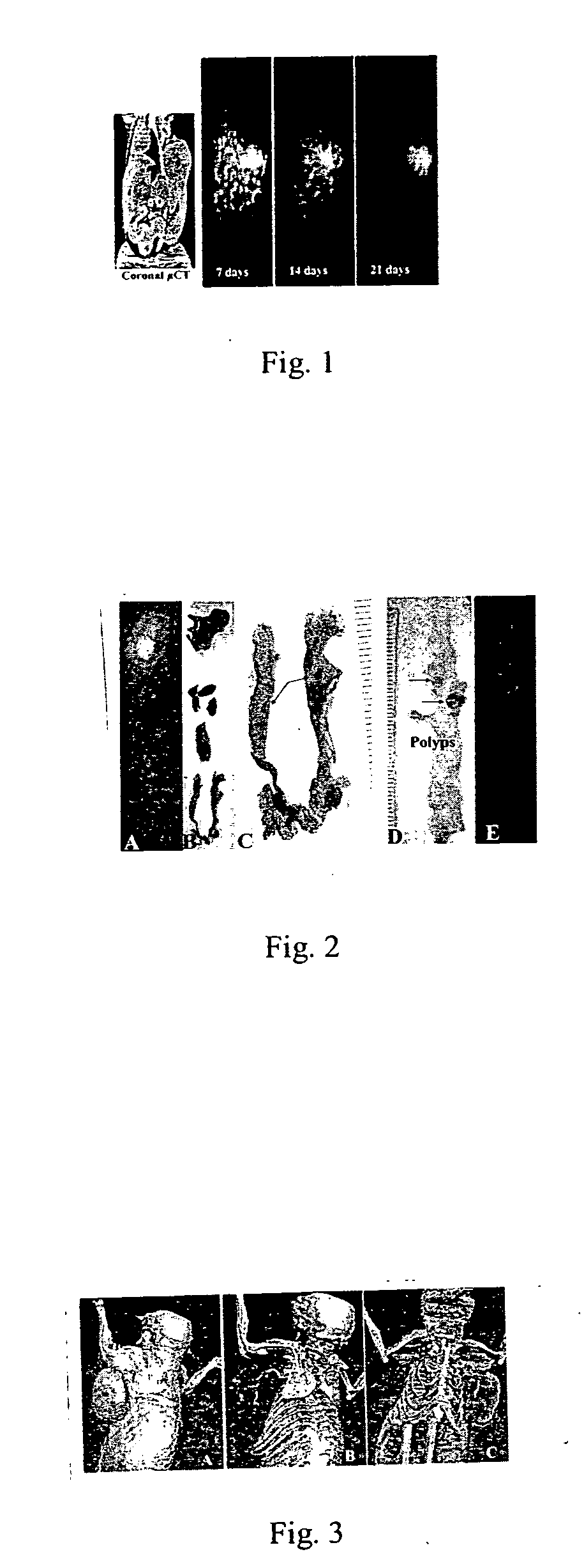
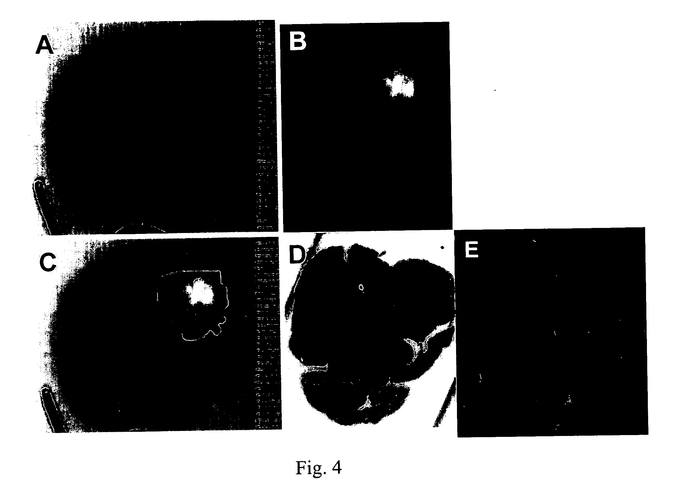
[0027] ApcMin / + Mouse Model: This model is comprised of mice carrying the Min allele of Apc (ApcMin / + mice) and the type of lesions that appear in these mice are molecularly and histologically similar to breast cancer in humans. This model offers specific advantages over xenograft models in that female ApcMin / + mice are predisposed to developing mammary hyperplasias and carcinomas and intestinal adenomas. On the C57BL6 / J genetic background, about 5% of untreated females will develop a mammary tumor by 100 days of age (Moser A R, et al. Proc. Natl. Acad. Sci. USA 90:8977-81, 1993). The incidence and multiplicity of the mammary lesions can be increased by a single dose of ethylnitrosourea (ENU), a direct acting alkylating agent. Treatment with ENU results in 90% of B6 ApcMin / + females developing an average of 3 mammary squamous cell carcinomas (SCC), but...
example 2
Imaging of Intracranial Gliomas Using Radioiodinated NM404
Materials and Methods
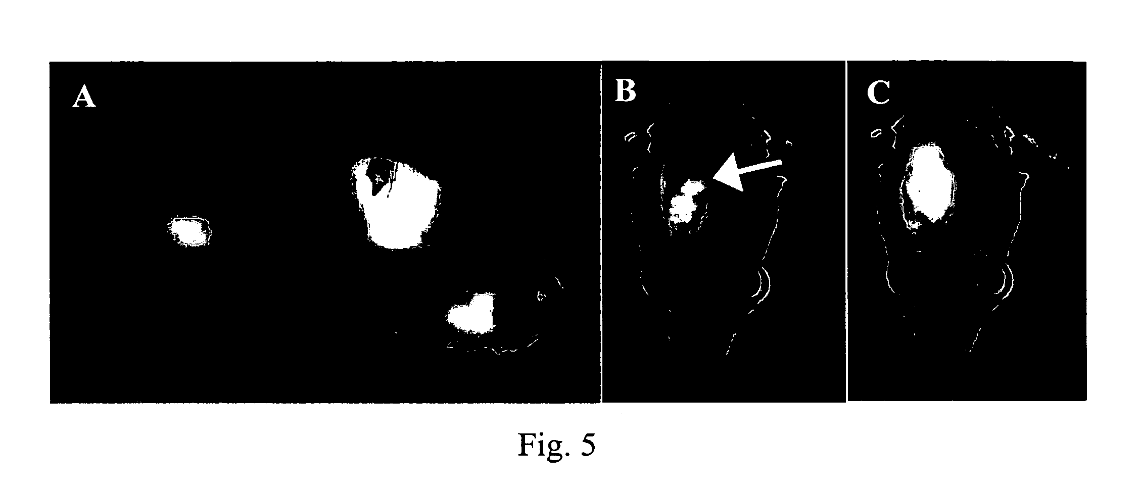
[0032] Glioma tumor model: All animals were housed and handled in accordance with the University of Wisconsin Research Animal Resources Center guidelines. Rat C6 glioma cells were propagated in DMEM medium (Life Technologies, Gaithersburg, Md.) supplemented with 10% heat-inactivated FBS (BioWhittaker, Walkersville, MD), 100 U / ml penicillin-G, 100 μg / ml streptomycin, and 0.01 M hepes (Life Technologies, Gaithersburg, Md.). Intracranial tumor implantation was performed as described in Badie B, et al. J. Neuroimmunol. 133:39-45, 2002. Briefly, 1×10⁶ C6 cells were resuspended in 5 μl of 1.2% methylcellulose and injected into the frontal lobes of anesthetized female Wistar rats (Harlan, Indianapolis, Ind.). Sham-operated animals received intracranial injections of an equal volume of methylcellulose without tumor cells.
[0033] Imaging studies: Ten days after implantation rats were screened for the presence o...
example 3
[0038] MicroPET Evaluation of 124I labeled NM404 in a Rat CNS-1 Brain Tumor Model
Materials and Methods
[0039] NM404 was radiolabeled with 124I in excellent radiochemical yield (>60% isolated yield, >99% purity) via isotope exchange reaction of stable NM404 with sodium-iodide (Eastern Isotopes, Sterling Va.). Following preparative HPLC purification 124I-NM404 was solubilized in 2% Tween-20 and filtered (0.22 micron). NM404 (130-200 μCi in 0.1 ml) was injected (i.v., tail vein) into 6 rats with CNS-1 brain tumor xenografts. MicroPET images (Concorde Microsystems-P4, 30 min acquisition) were acquired immediately after and at 6 h, 24 h, and 96 h post injection. Contrast-enhanced MRI images were obtained immediately following the final PET scan and PET / MRI images were manually fused using Amira (V3.1, TGS, Inc). Rats were euthanized and brain tissue subjected to histopathologic analysis.
Results
[0040]124 I-NM404 showed no tumor uptake within 30 minutes of injection and respectable tu...
PUM
 Abstract
Description
Claims
Application Information
 - R&D
- Intellectual Property
- Life Sciences
- Materials
- Tech Scout
- Unparalleled Data Quality
- Higher Quality Content
- 60% Fewer Hallucinations
Browse by: Latest US Patents, China's latest patents, Technical Efficacy Thesaurus, Application Domain, Technology Topic, Popular Technical Reports.
© 2025 PatSnap. All rights reserved.Legal|Privacy policy|Modern Slavery Act Transparency Statement|Sitemap|About US| Contact US: help@patsnap.com


