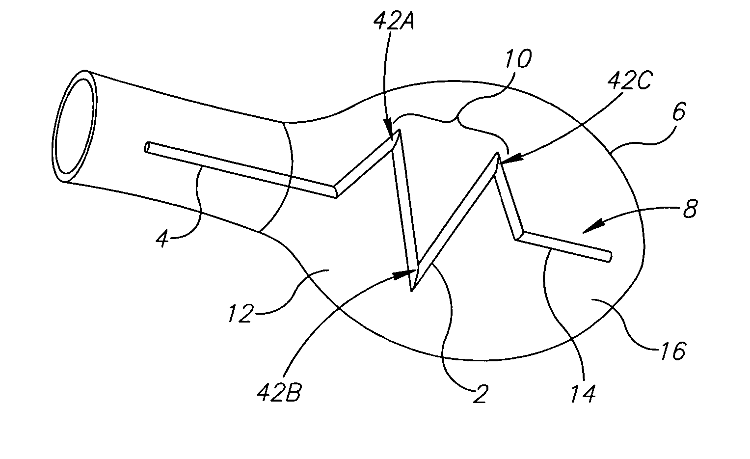
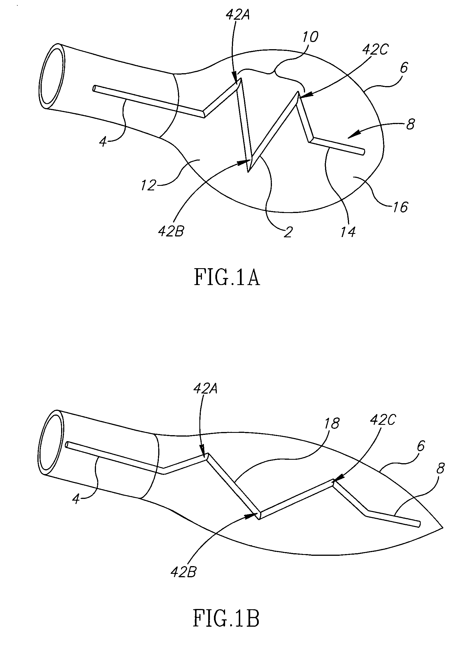
[0032] An aspect of some embodiments of the present invention relates to a flexible cardiac catheter with an intraventricular portion characterized by a large transverse proximal aspect. Optionally, the transverse proximal aspect of the intraventricular portion is larger than a transverse distal aspect of the intraventricular aspect. In an exemplary embodiment of the invention, employing a catheter with an increased proximal aspect reduces the chance of unwanted catheter ejection during ventriculography. In an exemplary embodiment of the invention, the transverse proximal aspect of the intraventricular portion is larger than a
heart valve through which the catheter has been inserted but smaller than the ventricular base. Optionally, this prevents unwanted ejection of the catheter through the valve. The valve may optionally be an aortic or
mitral valve. Optionally, this reduces mechanical stimulation of the myocardium which might lead to arrhythmia,
[0034] In an exemplary embodiment of the invention, the catheter includes a portion characterized by an undulating curve so that the catheter is substantially flat. In an exemplary embodiment of the invention, the catheter includes a portion characterized by one or more
helical turns so that the catheter is three dimensional. Optionally, the curvature conforms to ventricular geometry during
diastole. In an exemplary embodiment of the invention, the catheter does not contact an inner surface of the myocardium during
diastole. In an exemplary embodiment of the invention, the curved portions of the catheter are more flexible than intervening portions in order to help reduce
mechanical irritation of a
ventricular wall during
ventricular contraction. Optionally, flexibility of the catheter permits conformation of the catheter to a reduced systolic
ventricular cavity size and reduces
mechanical irritation. Alternatively or additionally, a portion of the catheter outside the
ventricle may include a flexible joint capable of conforming to a changing angle between the intraventricular portion of the catheter and the extra ventricular (e.g. aortic) portion of the catheter. Optionally, this feature further reduces
mechanical irritation and contributes to a reduction in arrhythmia.
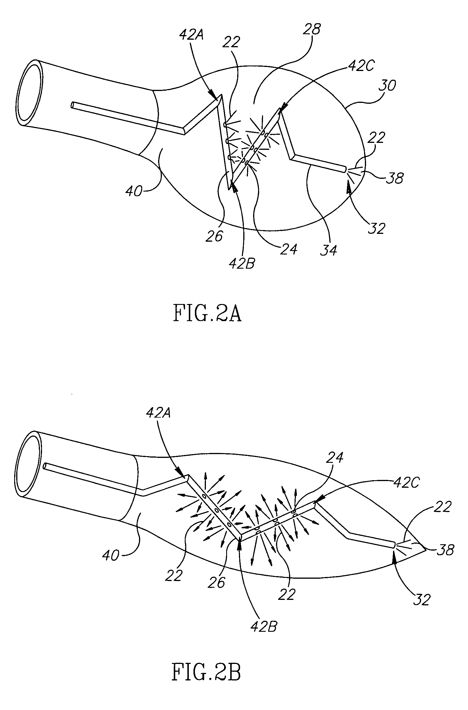
[0036] An aspect of some embodiments of the present invention relates to a method to reduce the amount of contrast material required for cardiac ventriculography by providing a large contrast exit area along the length of a curved catheter. Optionally, the contrast exit area is positioned and / or oriented to deliver contrast material to the
central region of the ventricle. Optionally, a large contrast exit area is achieved by a large number of ports and / or ports with a large cross-sectional area. In an exemplary embodiment of the invention, a large number of contrast material ports permits adequate
image quality with a reduced amount of contrast material. Optionally, the large contrast exit area reduces a resistance pressure for injection and / or provides efficient distribution of contrast material and / or permits use of more flexible materials in catheter construction. In an exemplary embodiment of the invention, at least 8, optionally 10, optionally 12, optionally 16 or more contrast material ports are provided. In an exemplary embodiment of the invention, the large contrast exit area eliminates the need for connection to a mechanical contrast material pump. Optionally this reduces the
procedure time and / or the risk of blood
clot formation.
[0037] An aspect of some embodiments of the present invention relates a method to reduce the
irritation of the
ventricular wall during ventriculography. Optionally, a catheter configuration which conforms to cardiac geometry reduces the
irritation. Optionally, cardiac geometry refers to ventricular geometry. In an exemplary embodiment of the invention, conformation to ventricular geometry is achieved during
diastole and
systole via use of a flexible catheter. Optionally, a catheter configuration with a transverse proximal aspect that is larger than a transverse distal aspect provides a desired degree of conformation to ventricular geometry. Optionally, cardiac geometry includes the aortic ventricular junction. In an exemplary embodiment of the invention, conformation to the dynamic angular configuration of this junction may be achieved by introducing at least one extraventricular flex point on the catheter. Alternatively or additionally, a
fixed angle in a portion of the catheter deployed in the
aorta aids in correct positioning of a portion of the catheter deployed within the
aorta. In an exemplary embodiment of the invention, an envelope of the catheter may conform the ventricular envelope during diastole and / or or
systole.
[0062] In an exemplary embodiment of the invention, there is provided a method of reducing mechanical
irritation of a
ventricular wall during a
medical diagnostic procedure. The method includes employing a catheter having an operative head which responds to an applied contractile force with a
low resistance to deliver contrast material for the procedure. Optionally, said catheter is sufficiently elastic to alternate between a first contracted conformation and a second extended conformation in accord with a degree of externally applied force.


