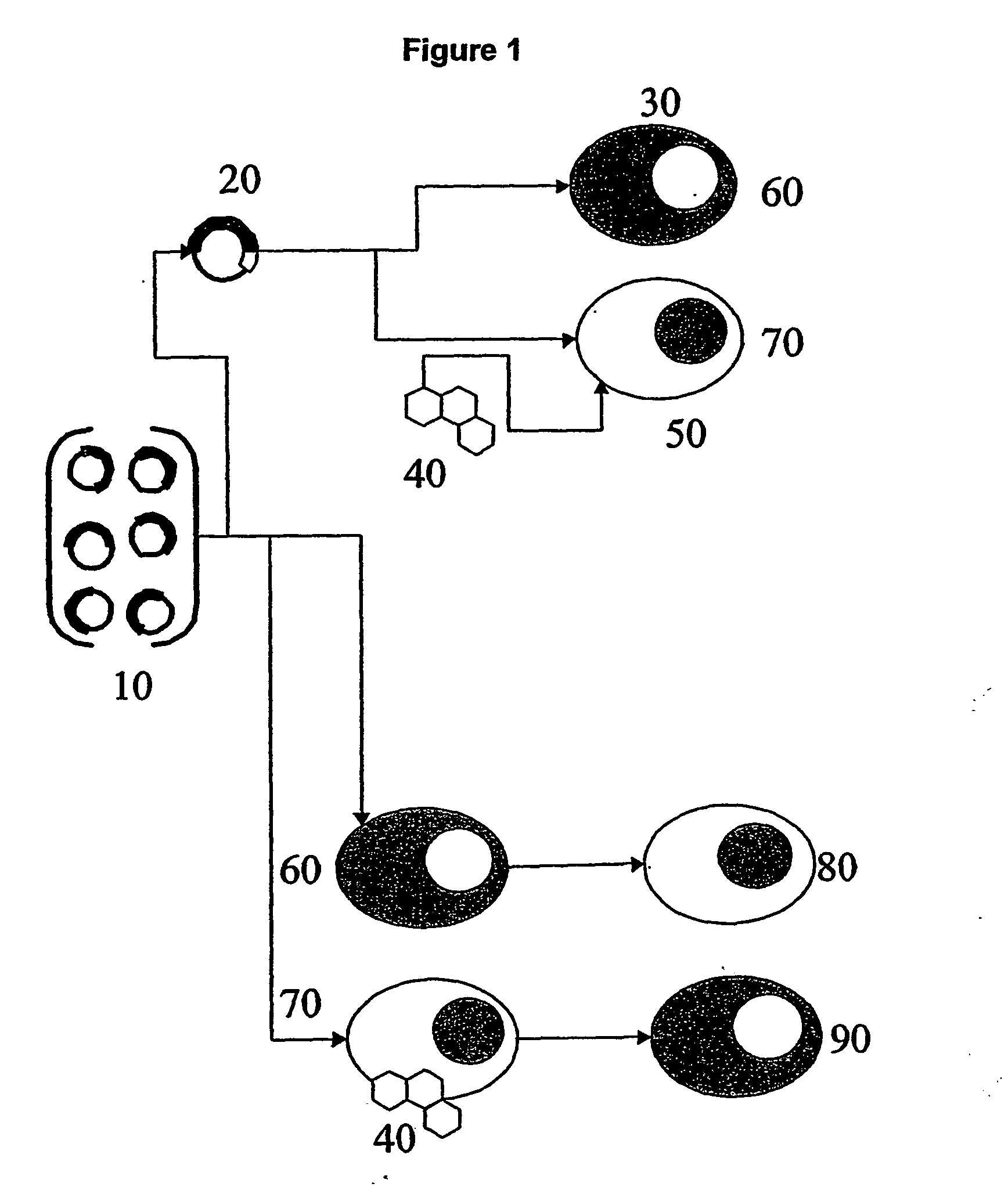
Functional screening method
a functional screening and genomic technology, applied in the field of high-throughput functional screening methods, can solve the problems of limited scope, limited coverage of biological events, and limited function assignment,
- Summary
- Abstract
- Description
- Claims
- Application Information
AI Technical Summary
Problems solved by technology
Method used
Image

Examples
specific examples
Example 1
[0082] A collection of cDNAs (Invitrogen & Image Consortium, Table 2) were prepared for expression as cDNA-EGFP fusion proteins by inserting cDNA sequences into the multiple cloning site of pCORON 1000-EGFP-N2 and pCORON1000-EGFP-C1 expression vectors (Amersham Biosciences) using standard molecular cloning techniques (Molecular Cloning, Sambrook & Russell, Cold Spring Harbour Press 2001). These vectors direct the expression of fusion proteins comprising the protein encoded by the inserted cDNA sequence fused at their amino and carboxy termini to EGFP in mammalian cells under the control of a constitutively active CMV promoter.
[0083] Expression vectors encoding cDNA-EGFP indicators were transiently transfected into HeLa cells growing in wells of 96 well microtitre plates by chemically mediated transfection (Fugene, Roche) and cells incubated under standard growth conditions for 24 hours to permit synthesis of indicator fusion proteins. Cells were subsequently stained with ...
example 2
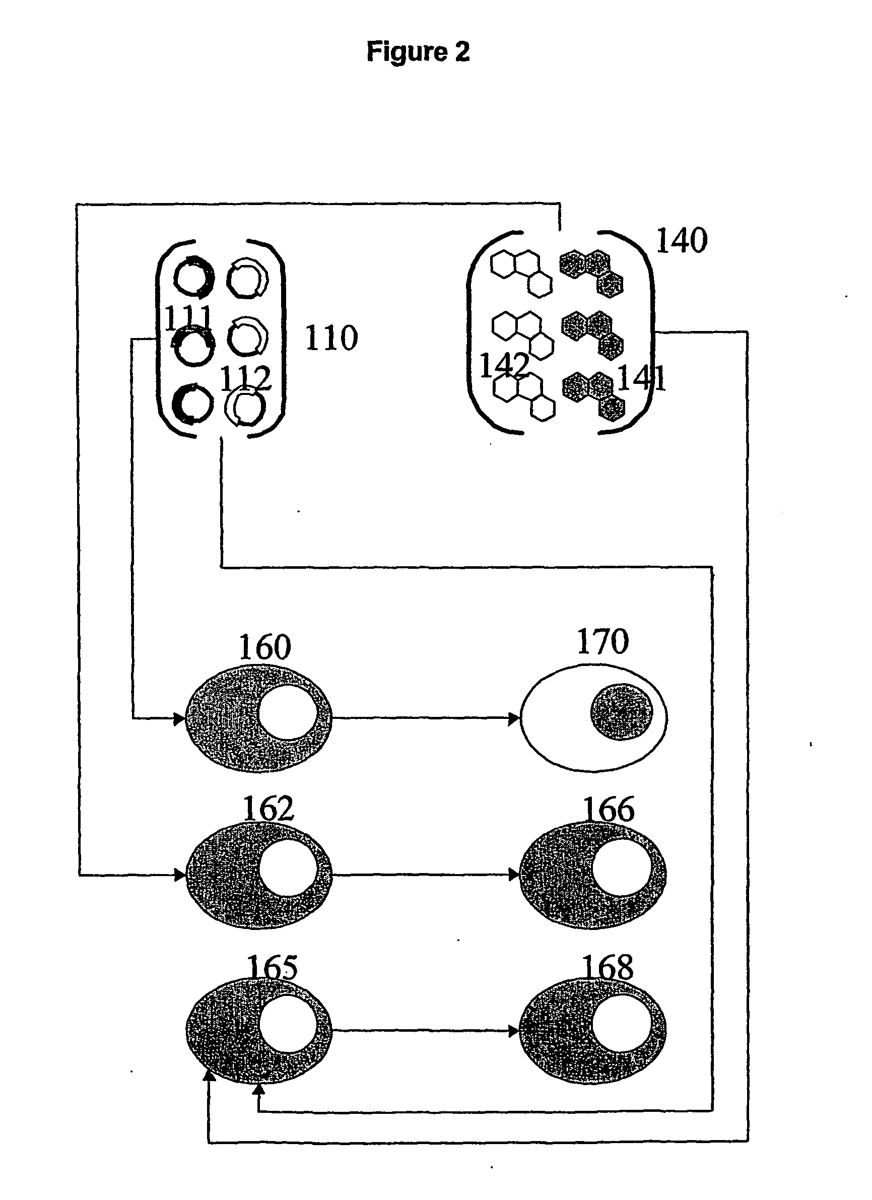
[0085] Indicator proteins derived from a range of cDNAs as described for Example 1 were transfected into HeLa cells and allowed to express for 24 hours. Following expression, cells were transferred into serum-free media for 2 hours to allow effects of stimuli from serum factors such as cortisol to decay. Cells were stained with DRAQ 5, imaged as described in Example 1, returned to complete media and then exposed to 1 μM dexamethasone (a synthetic glucocorticoid agonist) or 1 μM staurosporine (kinase inhibitor and apoptosis inducer) for 5 minutes followed by repeat imaging. Image data were analysed using a nuclear trafficking algorithm (Amersham Biosciences; (cf. Adie et al. (2001) ‘The pharmacological characterisation of a GPCR using pH sensitive cyamine dyes on the LEADseeker Cell Analysis System’ Poster, Society for Biomolecular Screening Conference 10-13th Sep. 2001, Baltimore USA; Goodyer et al. (2001) ‘Screening of signalling events in live cells using novel GFP redistribution ...
example 3
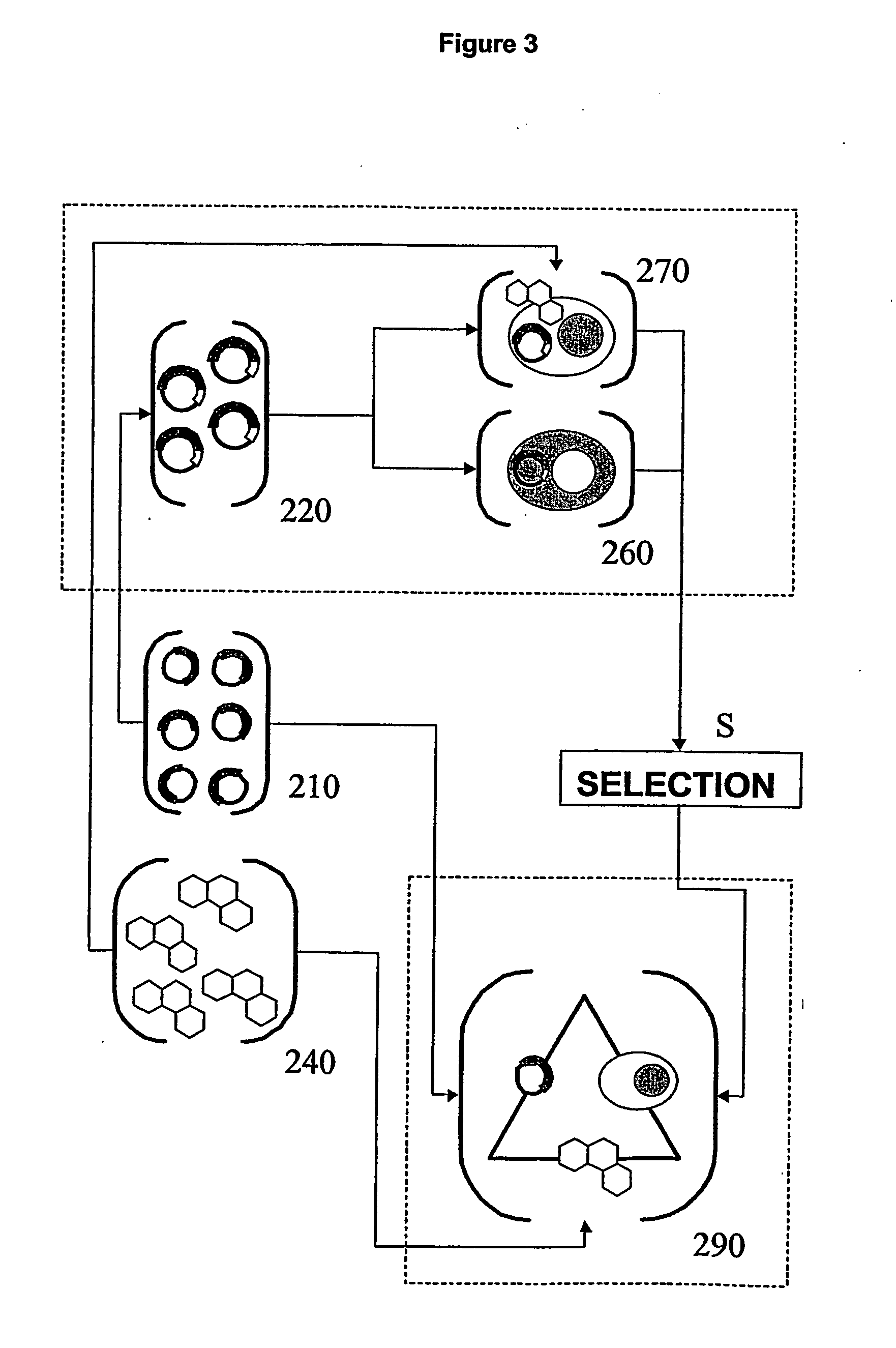
[0088] A further group of indicator proteins were transfected into HeLa cells and cells imaged before and after exposure to staurosporine as described in Example 2. Images were analysed with a further two IN Cell Analyzer algorithms, Granularity and Membrane Spot (Amersham Biosciences) (cf. Adie et al. (2001) ‘The pharmacological characterisation of a GPCR using pH sensitive cyamine dyes on the LEADseeker Cell Analysis System’ Poster, Society for Biomolecular Screening Conference 10-13th Sep. 2001, Baltimore USA; Goodyer et al. (2001) ‘Screening of signalling events in live cells using novel GFP redistribution assays’ Poster, Society for Biomolecular Screening Conference 10-13th Sep. 2001). These algorithms return results which quantitate fluorescence in degrees of granularity (i.e. low value indicates uniform distribution, high value indicates punctate distribution) and in terms of membrane localisation. Consequently these algorithms are suitable for examining indicators which no n...
PUM
| Property | Measurement | Unit |
|---|---|---|
| optical | aaaaa | aaaaa |
| fluorescent | aaaaa | aaaaa |
| Fluorescent | aaaaa | aaaaa |
Abstract
Description
Claims
Application Information
 - R&D
- Intellectual Property
- Life Sciences
- Materials
- Tech Scout
- Unparalleled Data Quality
- Higher Quality Content
- 60% Fewer Hallucinations
Browse by: Latest US Patents, China's latest patents, Technical Efficacy Thesaurus, Application Domain, Technology Topic, Popular Technical Reports.
© 2025 PatSnap. All rights reserved.Legal|Privacy policy|Modern Slavery Act Transparency Statement|Sitemap|About US| Contact US: help@patsnap.com