Embryonic epithelial cells
a technology epithelial cells, applied in the field of embryonic stem cells, can solve the problems that the effect of biomaterials on stem cell behavior has not been studied in great detail
- Summary
- Abstract
- Description
- Claims
- Application Information
AI Technical Summary
Problems solved by technology
Method used
Image
Examples
example 1
Production of a Polymer Array
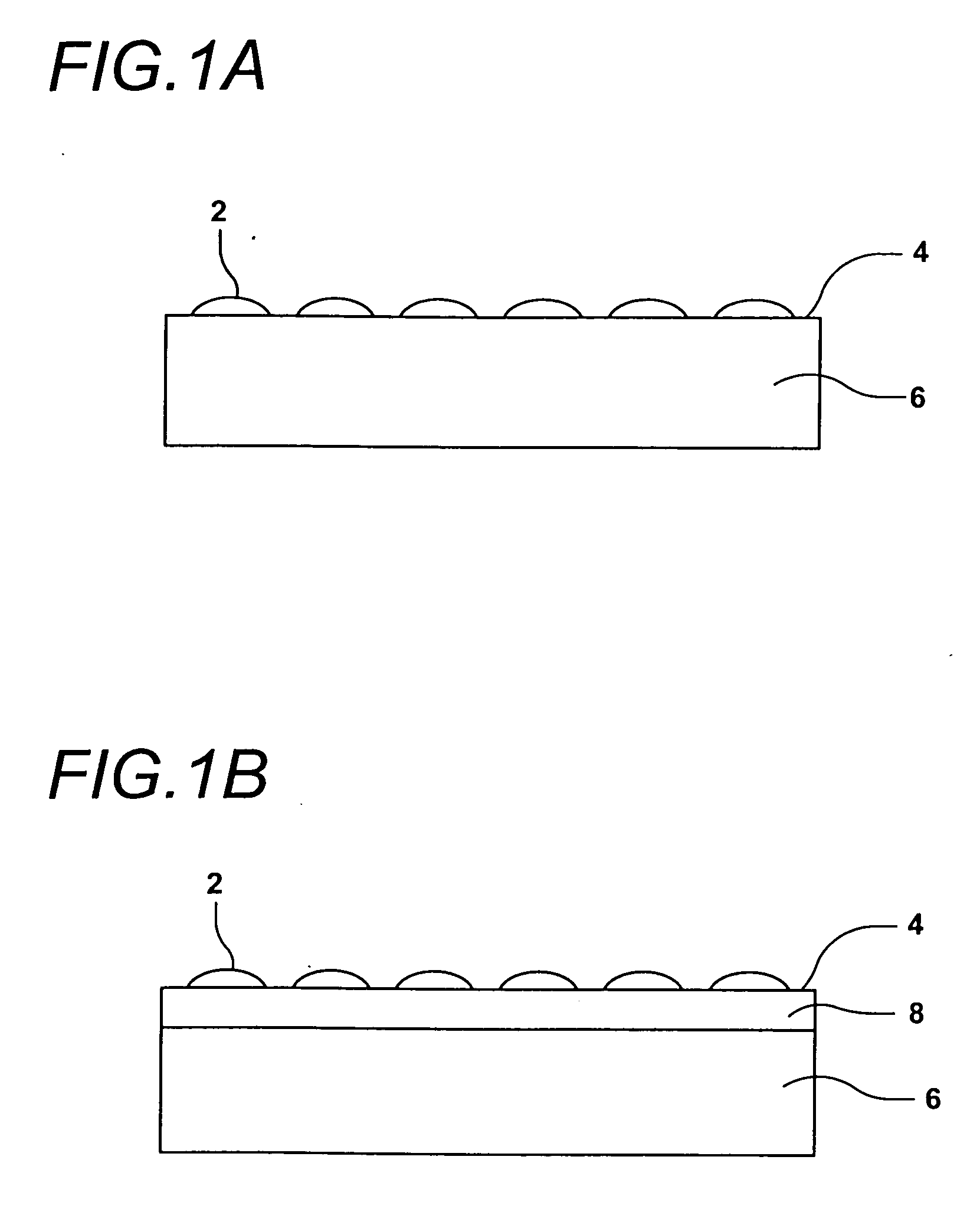
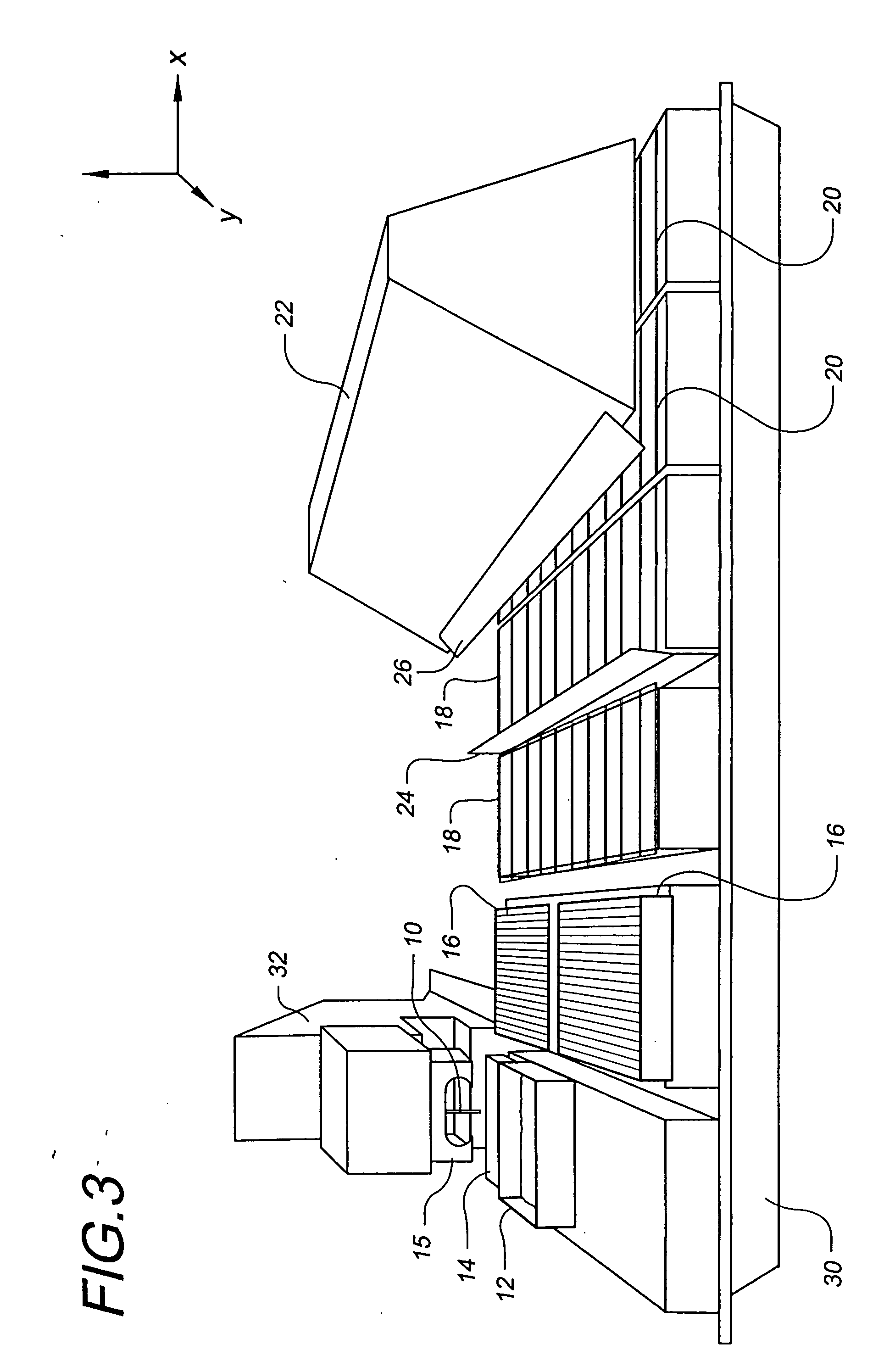
[0109] The use of robotic fluid handling for the production of DNA, protein, and small molecule microarrays is well defined (G. MacBeath, et al., Journal of the American Chemical Society 121, 7967-7968 (1999); G. MacBeath, et al., Science 289, 1760-1763 (2000); M. Schena, et al., Science 270, 467-470 (1995)). However, the deposition of structurally diverse acrylate monomers to produce a uniform, cell-compatible polymer microarray required significant modification of existing robotic technology. First, some acrylate monomers are viscous, affecting all aspects of monomer printing including pre-printing pin priming, fluid ejection at printing, and pin washing. Another problem unique to these arrays is that the ordinary sensitivity of radical polymerization to oxygen inhibition is particularly evident at small volumes. Consequently, we performed our printing in an atmosphere of humid argon with oxygen present at less than 0.1%. Humidity helps minimize faile...
example 2
Cell Culture
[0113] H9 cells (Thomson, J. A., et al., “Embryonic stem cells lines derived from human blastocysts”, Science 282, 1145-1147 (1998)) were grown as described in Spradling, A., et al., “Stem cells find their niche”, Nature 414, 98-104 (2001), the entire contents of which are incorporated herein by reference. C2C12 cells were grown as described in Yaffee, D. & Saxel, O., “Serial passaging and differentiation of myogenic cells isolated from dystrophic mouse muscle”, Nature 270, 725-7 (1977). Specifically, hES cells (H9 clone) were grown on mouse embryo fibroblasts (Cell Essential) in KnockOut Medium (Gibco-BRL, Gaithersburg, Md.), a modified version of Dulbeco's modified Eagle's medium optimized for ES cells (Itskovitz-Eldor, et. al., (2000) Mol. Med. 6, 88-95, the contents of which are incorporated herein by reference). Tissue cover plates were covered with 0.1% gelatin (Sigma). Culture were grown in 5% CO2 and were routinely passaged every 5-6 days after disaggregating wi...
example 3
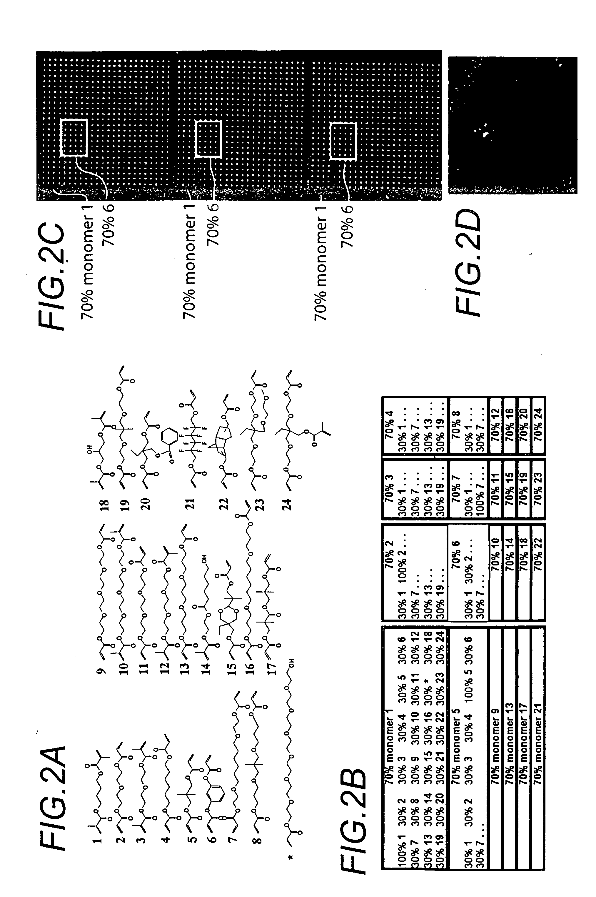
[0114] Chips were washed, fixed in 4% paraformaldehyde for 8 minutes, blocked with 10% goat serum (Zymed, San Francisco, Calif.) and permeablized with 0.2% triton X-100 for 30 minutes. Primary antibodies, Ms anti-Cytokeratin 7, Ms anti-Myogenin (Dako, Carpinteria, Calif.), Rb anti-Vimentin (Biomeda, Foster City, Calif.) in PBS with 3% goat serum were incubated on the chips for 1 hr. Chips were washed 3 times in 1% goat serum PBS. A mixture of Goat anti-Ms Alexa 555, Goat anti Rb Alexa, and SytoX24 (Molecular Probes, Eugene, Oreg.) were diluted into 3% goat serum PBS and incubated on the chips for 1 hr. Slides were washed 3 times in 1% goat serum PBS and dipped in 0.5 mM Tris Cl pH 7.5 to remove salt, and air dried immediately prior to scanning. Slides were then scanned using an Arrayworx autoloader scanner (API, Issaquah, Wash.) (FIG. 2).
PUM
| Property | Measurement | Unit |
|---|---|---|
| volume | aaaaa | aaaaa |
| volumes | aaaaa | aaaaa |
| volumes | aaaaa | aaaaa |
Abstract
Description
Claims
Application Information
 - R&D
- Intellectual Property
- Life Sciences
- Materials
- Tech Scout
- Unparalleled Data Quality
- Higher Quality Content
- 60% Fewer Hallucinations
Browse by: Latest US Patents, China's latest patents, Technical Efficacy Thesaurus, Application Domain, Technology Topic, Popular Technical Reports.
© 2025 PatSnap. All rights reserved.Legal|Privacy policy|Modern Slavery Act Transparency Statement|Sitemap|About US| Contact US: help@patsnap.com


