Inhibition of inflammatory cytokine production by cholinergic agonists and vagus nerve stimulation
- Summary
- Abstract
- Description
- Claims
- Application Information
AI Technical Summary
Benefits of technology
Problems solved by technology
Method used
Image
Examples
example 1
Cholinergic Agonists Inhibit Release of Proinflammatory Cytokines from Macrophages
1. Materials and Methods
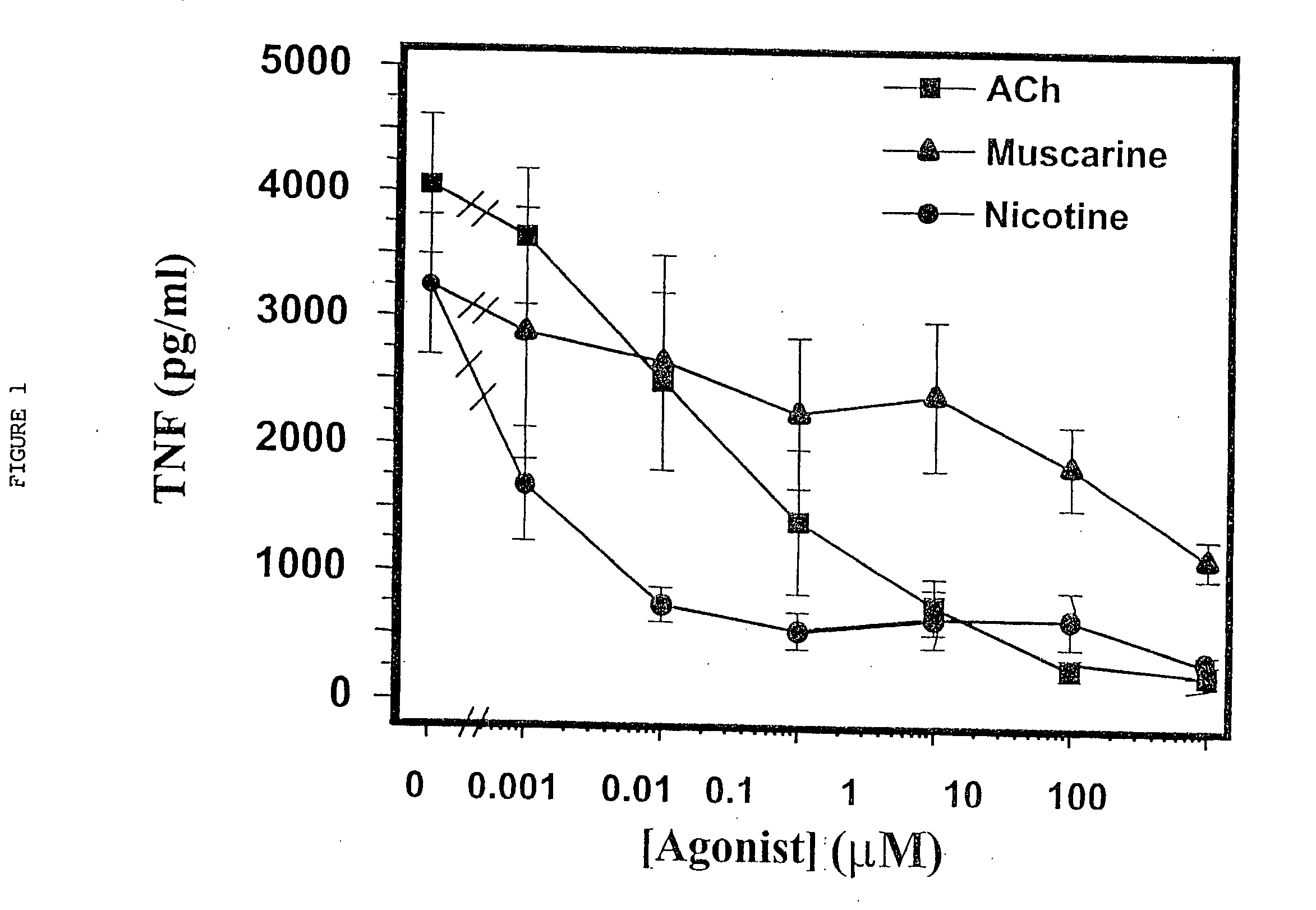
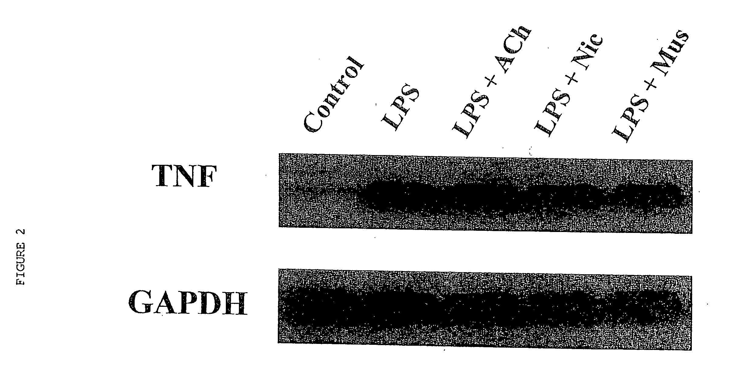
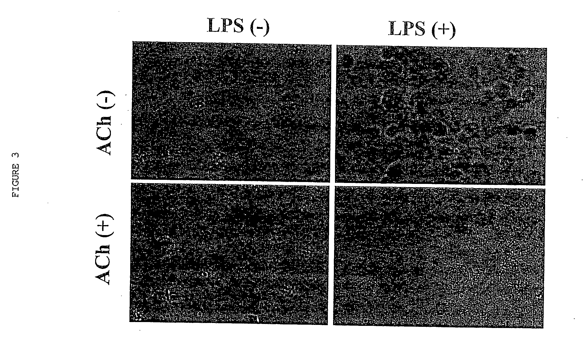
[0064] Human macrophage cultures were prepared as follows. Buffy coats were collected from the blood of healthy individual donors to the Long Island Blood Bank Services (Melville, N.Y.). Primary blood mononuclear cells were isolated by density-gradient centrifugation through Ficoll / Hypaque (Pharmacia, N.J.), suspended (8.times.10.sup.6 cells / ml) in RPMI 1640 medium supplemented with 10% heat inactivated human serum (Gemini Bio-Products, Inc., Calabasas, Calif.), and seeded in flasks (PRIMARIA; Beckton and Dickinson Labware, Franklin Lakes, N.J.). After incubation for 2 hours at 37.degree. C., adherent cells were washed extensively, treated briefly with 10 mM EDTA, detached, resuspended (10.sup.6 cells / ml) in RPMI medium (10% human serum), supplemented with human macrophage colony stimulating factor (MCSF; Sigma Chemical Co., St. Louis, Mo.; 2 ng / ml), and seeded onto 24-well t...
example 2
Inhibition of Endotoxic Shock by Stimulation of Efferent Vagus Nerve Fibers
[0079] To determine whether direct stimulation of efferent vagus nerve activity might suppress the systemic inflammatory response to endotoxin, adult male Lewis rats were subjected to bilateral cervical vagotomy, or a comparable sham surgical procedure in which the vagus nerve was isolated but not transected. Efferent vagus nerve activity was stimulated in vagotomized rats by application of constant voltage stimuli to the distal end of the divided vagus nerve 10 min before and again 10 min after the administration of a lethal LPS dose (15 mg / kg, i.v.). An animal model of endotoxic shock was utilized in these experiments. Adult male Lewis rats (280-300 g, Charles River Laboratories, Wilmington, Mass.) were housed at 22.degree. C. on a 12 h light / dark cycle. All animal experiments were performed in accordance with the National Institute of Health Guidelines under the protocols approved by the Institutional Ani...
example 3
Stimulation of Intact Vagus Nerve Attenuates Endotoxic Shock
[0089] Experiments were conducted to determine whether the inhibition of inflammatory cytokine cascades by efferent vagus nerve stimulation is effective by stimulation of an intact vagus nerve. Stimulation of left and right vagus nerves were also compared.
[0090] The vagus nerves of anesthetized rats were exposed, and the left common iliac arteries were cannulated to monitor blood pressure and heart rate. Endotoxin (E. coli 0111:B4; Sigma) was administered at a lethal dose (60 mg / kg). In treated animals, either the left or the right intact vagus nerve was stimulated with constant voltage (5V or 1V, 2 ms, 1 Hz) for a total of 20 min., beginning 10 min. before and continuing 10 min. after LPS injection. Blood pressure and heart rate were through the use of a Bio-Pac M100 computer-assisted acquisition system. FIGS. 12-14 show the results of these experiments.
[0091] As shown in FIG. 12, within minutes after LPS injection, the...
PUM
 Abstract
Description
Claims
Application Information
 - R&D
- Intellectual Property
- Life Sciences
- Materials
- Tech Scout
- Unparalleled Data Quality
- Higher Quality Content
- 60% Fewer Hallucinations
Browse by: Latest US Patents, China's latest patents, Technical Efficacy Thesaurus, Application Domain, Technology Topic, Popular Technical Reports.
© 2025 PatSnap. All rights reserved.Legal|Privacy policy|Modern Slavery Act Transparency Statement|Sitemap|About US| Contact US: help@patsnap.com


