Method and device for biochemical detection and analysis of subcellular compartments from a single cell
- Summary
- Abstract
- Description
- Claims
- Application Information
AI Technical Summary
Benefits of technology
Problems solved by technology
Method used
Image
Examples
example 1
On-Chip Integration
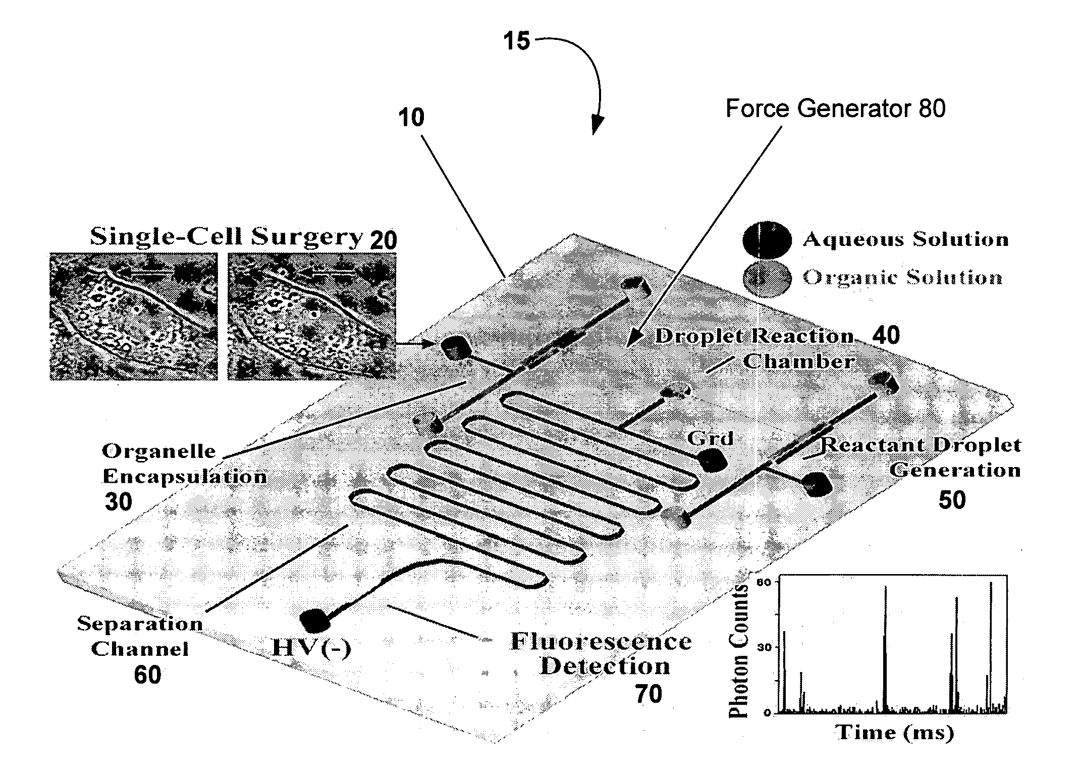
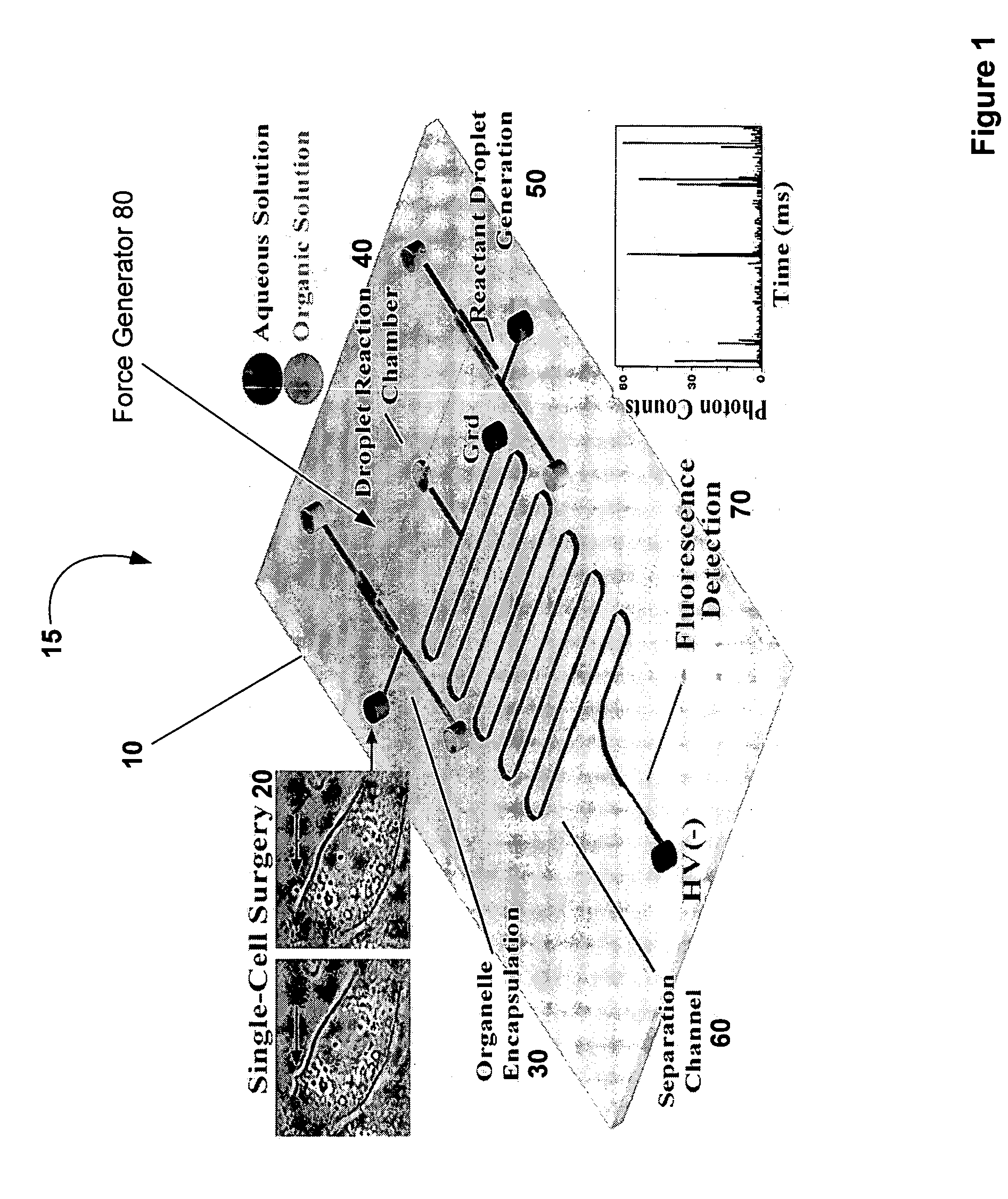
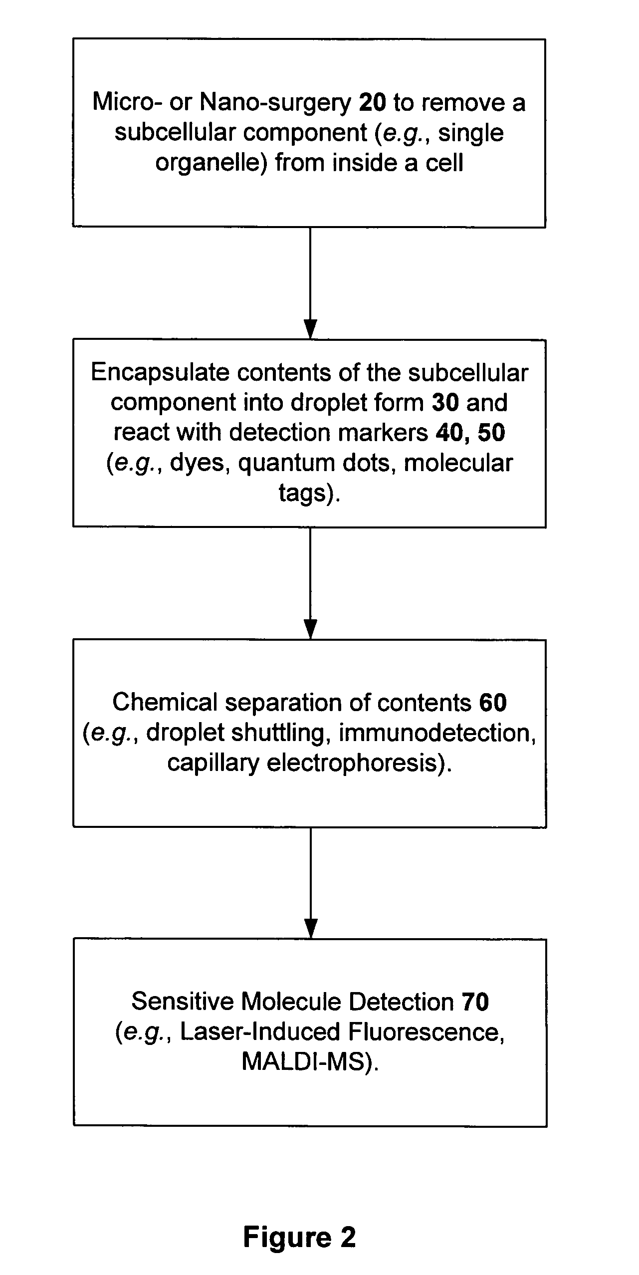
The interface combination of these techniques was performed on a single chip 15 as illustrated in FIG. 1 (e.g., fabricated in a substrate 10, for example, polydimethylsiloxane, polymethylmethacrylate, glass, silicon, polyester, and / or other materials, and combinations thereof) with micro- and nano-fluidic channels, in which single-cell surgery 20 was performed on the substrate 10, followed by the in-channel droplet generation 50 along with the microand / or nano-scale chemical reaction 40, with each component of the reacted mixture subsequently separated by CE in microchannels 60. In the case of laser-induced fluorescence detection, the separated components are detected within the constricted region of the micro- and / or nano-channel 70. In the case of mass spectrometric detection, the separated components or the intact organelles are ionized into the gas phase for mass spectrometric detection. The characterization of each process step is described in sections fol...
example 2
Single-Cell Micro- or Nano-Surgery and Micro- or Nano-Scale Chemical Reaction
To isolate single subcellular compartments by single cell nano-surgery 20, optical trapping was used as a force generator 80 to first manipulate and transport the identified organelles close to the cell membrane (FIG. 7A). Optical trapping has been used in a wide range of applications, from the manipulation of micro- and nano-meter sized particles to single cells and subcellular structures. A single nanometer scale organelle was then moved from the cellular interior to a location adjacent to the membrane surface (FIGS. 7B and 7C, arrow). Once parked at the membrane, the organelle is transported across the cell membrane. A focused ultraviolet (UV) or near infrared (IR) laser (FIGS. 7D-7G), was used to “open” a small nanometer-sized patch on the cell membrane to isolate the organelle from the cell for chemical analysis. Because the cell is a heavily compartmentalized structure, this approach is applicable ...
example 3
Sensitive Detection of Single Fluorescent Molecules
The detection 70 of single fluorescent molecules can be achieved with an excellent signal-to-noise ratio, and has become more facile and routine with improved detectors and optics. The detection volume of a single-molecule confocal fluorescence microscope was defined latitudinally by the laser focus and longitudinally by the pinhole present at the image plane. This far-field method for single-molecule detection shows excellent signal-to-noise ratio at ˜1-ms photon integration time, which is illustrated in FIG. 9. FIG. 9 shows a photon trace showing the detection of single carboxyrhodamine 6G molecule diffusing in solution, in which each spike signals the presence of a single dye molecule within the probe volume. One criterion to achieve such high signal-to-noise ratio is to minimize the laser probe volume by using a diffraction limited laser focus, which is ˜0.5 Hm in diameter and 2 μm in height. This small detection volume ensur...
PUM
| Property | Measurement | Unit |
|---|---|---|
| Volume | aaaaa | aaaaa |
| Volume | aaaaa | aaaaa |
| Force | aaaaa | aaaaa |
Abstract
Description
Claims
Application Information
 - R&D Engineer
- R&D Manager
- IP Professional
- Industry Leading Data Capabilities
- Powerful AI technology
- Patent DNA Extraction
Browse by: Latest US Patents, China's latest patents, Technical Efficacy Thesaurus, Application Domain, Technology Topic, Popular Technical Reports.
© 2024 PatSnap. All rights reserved.Legal|Privacy policy|Modern Slavery Act Transparency Statement|Sitemap|About US| Contact US: help@patsnap.com


