Eureka AIR delivers breakthrough ideas for toughest innovation challenges, trusted by R&D personnel around the world.

Intramyocardial injection of autologous bone marrow

Inactive Publication Date: 2004-08-19
KOMOWSKI RAN +3
View PDF2 Cites 15 Cited by
  • Summary
  • Abstract
  • Description
  • Claims
  • Application Information

AI Technical Summary

Benefits of technology

[0006] It is also an object of this invention to provide a novel method of intramyocardial injection to enhance collateral blood vessel formation and tissue perfusion.
[0009] The bone marrow (BM) is a natural source of a broad spectrum of cytokines and cells that are involved in the control of angiogenic processes. It is therefore believed that the intramyocardial injection of autologous (A) BM, by taking advantage of the natural ability of these cells to secrete many angiogenic factors in a time-appropriate manner, provides an optimal intervention for achieving therapeutic collateral development in ischemic myocardium.
[0021] Because VEGF promoter activity is enhanced by HIF-1, this invention also includes the ex-vivo exposure of bone marrow cells in culture to hypoxia or other forms of energy, such as, for example, ultrasound, RF, or electromagnetic energy. This intervention increases VEGF and other gene expression. By this effect it may augment the capacity of bone marrow to stimulate angiogenesis.
[0025] Granulocyte-macrophage colony-stimulating factor (GM-CSF) and Granulocyte-Colony Stimulatory Factor (G-CSF) are stimulatory cytokines for monocyte maturation and are multipotent hematopoietic growth factors, which are utilized in clinical practice for various hematological pathologies such as depressed white blood cell count (i.e., leukopenia or granulocytopenia or monocytopenia) which occurs usually in response to immunosuppressive or chemotherapy treatment in cancer patients. GM-CSF has also been described as a multilineage growth factor that induces in vitro colony formation from erythroid burst-forming units, eosinophil colony-forming units (CSF), and multipotential (CSF), as well as from granulocyte-macrophage CSF and granulocyte CFU. (Bot F. J., Exp Hematol 1989, 17:292-5). Ex-vivo exposure to GM-CSF has been shown to induce rapid proliferation of CD-34+ progenitor cells (Egeland T. et al., Blood 1991; 78:3192-9.) These cells have the potential to differentiate into vascular endothelial cells and may naturally be involved in postnatal angiogenesis. In addition, GM-CSF carries multiple stimulatory effects on macrophage / monocyte proliferation, differentiation, motility and survival (reduced apoptotic rate). Consistent with the combined known effects on bone marrow derived endothelial progenitor cells and monocytes, it is another aspect of the invention to use GM-CSF as an adjunctive treatment to autologous bone marrow injections aimed to induce new blood vessel formation and differentiation in ischemic cardiovascular organs. Moreover, GM-CSF may further enhance therapeutic myocardial angiogenesis caused by bone marrow, by augmenting the effect of bone marrow, or by further stimulating, administered either in vivo or in vitro, bone marrow that is also being stimulated by agents such as HIF-1, EPAS1, hypoxia, or MCP-1.

Problems solved by technology

However, it is believed that complex interactions among several growth factor systems are probably necessary for the initiation and maintenance of new blood vessel formation.

Method used


Image

  • Intramyocardial injection of autologous bone marrow
  • Intramyocardial injection of autologous bone marrow

Examples


example 1

[0027] Effect of Bone Marrow Cultured Media on Endothelial Cell Proliferation

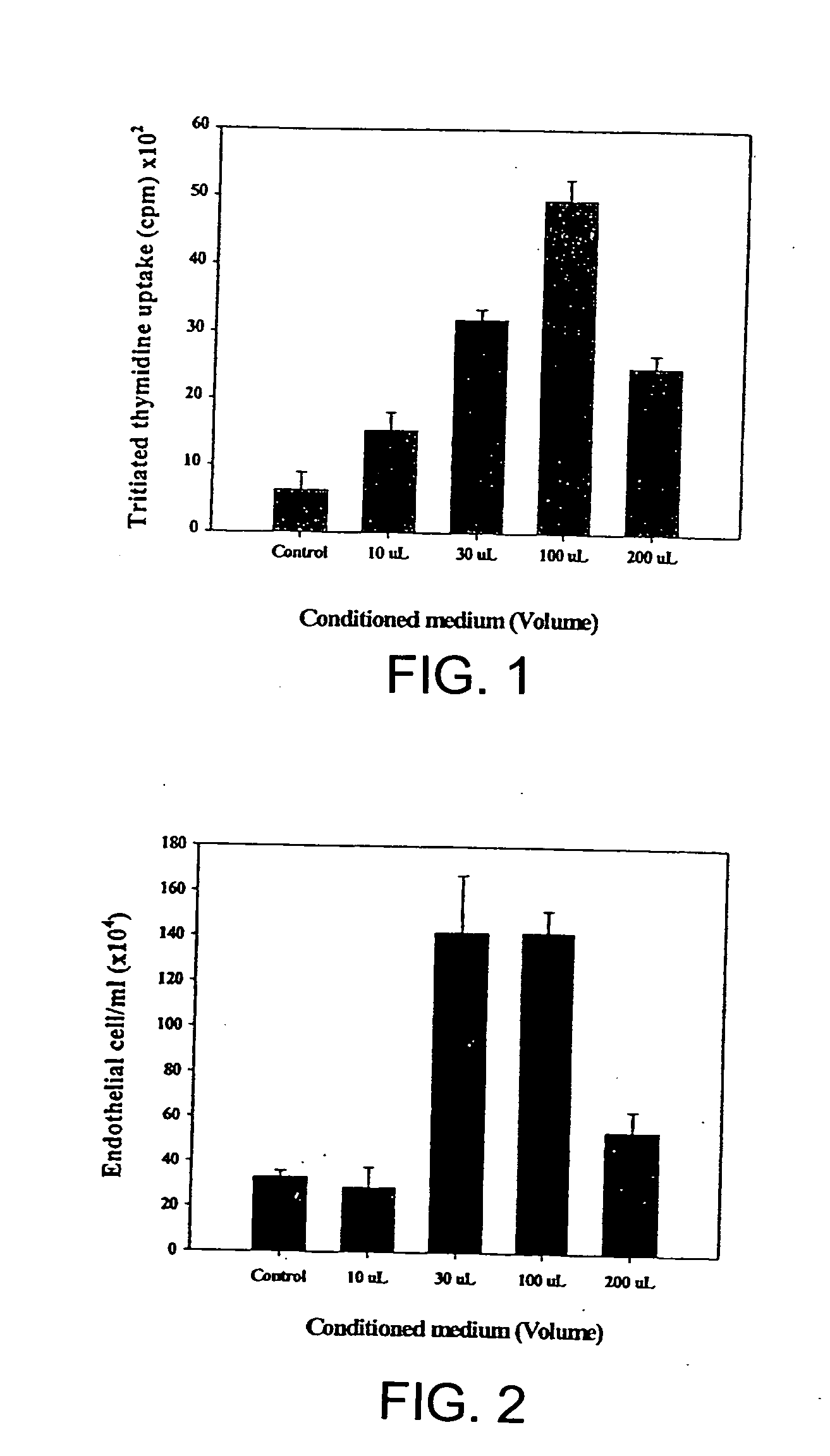
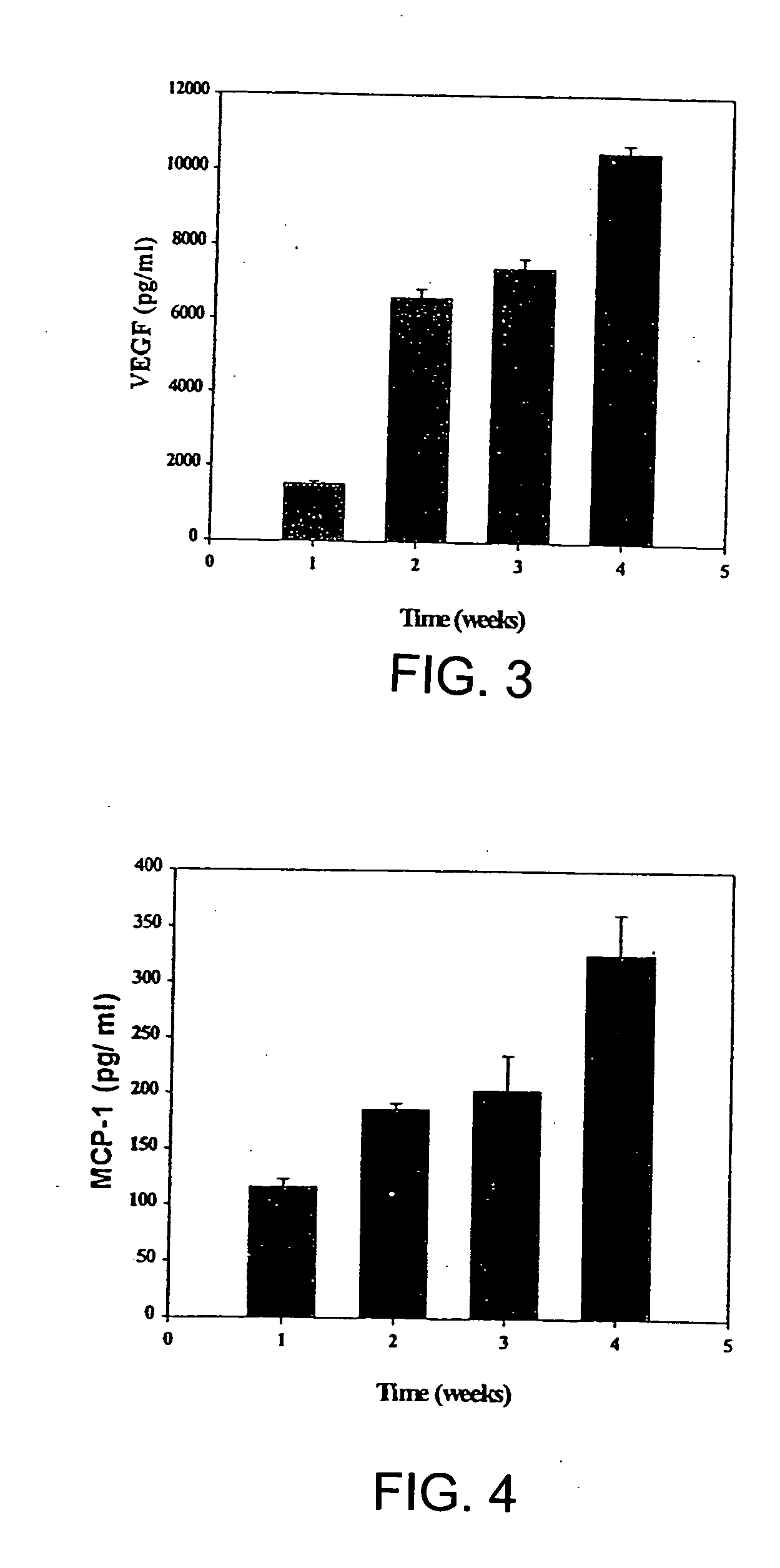
[0028] Studies were conducted to determine whether aspirated pig autologous bone marrow cells obtained secreted VEGF, a potent angiogenic factor, and MCP-1, which recently has been identified as an important angiogenic co-factor. Bone marrow was cultured in vitro for four weeks. The conditioned medium was added to cultured pig aortic endothelial cells (PAECs), and after four days proliferation was assessed. VEGF and MCP-1 levels in the conditioned medium were assayed using ELISA. During the four weeks in culture, BM cells secreted VEGF and MCP-1, such that their concentrations increased in a time-related manner. The resulting conditioned medium enhanced, in a dose-related manner, the proliferation of PAECs. The results indicate that BM cells are capable of secreting potent angiogenic cytokines such as VEGF and MCP-1 and of inducing proliferation of vascular endothelial cells.

[0029] Pig Bone Marrow Culture

[0...

example 2

[0045] Effects of Hypoxia on VEGF Secretion by Cultured Pig Bone Marrow Cells

[0046] It was demonstrated that hypoxia markedly increases the expression of VEGF by cultured bone marrow endothelial cells, results indicating that ex-vivo exposure to hypoxia, by increasing expression of hypoxia-inducible angiogenic factors, can further increase the collateral enhancing effect of bone marrow cells and its conditioned media to be injected in ischemic muscular tissue. Pig bone marrow was harvested and filtered sequentially using 300.mu. and 200.mu. stainless steel mesh filters. BMCs were then isolated by Ficoll-Hypaque gradient centrifugation and cultured at 33.degree. C. with 5% CO2 in T-75 culture flasks. When cells became confluent at about 7 days, they were split 1:3 by trypsinization. After 4 wks of culture, the BMCs were either exposed to hypoxic conditions placed in a chamber containing 1% oxygen) for 24 to 120 hrs, or maintained under normal conditions. The resulting conditioned med...

example 3

[0048] Effect of Bone Marrow Cultured Media on Endothelial Cell Tube Formation

[0049] It was demonstrated, using pig endothelial cells and vascular smooth muscle cells co-culture technique, that the conditioned medium of bone marrow cells induced the formation of structural vascular tubes in vitro. No such effect on vascular tube formation was observed without exposure to bone marrow conditioned medium. The results suggest that bone marrow cells and their secreted factors exert pro-angiogenic effects.


PUM

PropertyMeasurementUnit
Electrical conductivityaaaaaaaaaa
Cell proliferation rateaaaaaaaaaa

Abstract

A method of treating cardiac or myocardial conditions comprises the administration of an effective amount of autologous bone marrow. The bone marrow may optionally be stimulated and / or administered in combination with a pharmaceutical drug, protein, gene or other factor or therapy that may enhance bone marrow production of angiogenic growth factors and / or promote endothelial cell proliferation or migration or blood vessel formation.

Description

[0001] This application is directed to a method of injecting autologous bone marrow. More specifically, this invention is directed to intramyocardial injection of autologous bone marrow to enhance collateral blood vessel formation and tissue perfusion.[0002] The use of recombinant genes or growth-factors to enhance myocardial collateral blood vessel function may represent a new approach to the treatment of cardiovascular disease. Kornowski, R., et al., "Delivery strategies for therapeutic myocardial angiogenesis", Circulation 2000; 101:454-458. Proof of concept has been demonstrated in animal models of myocardial ischemia, and clinical trials are underway. Unger, E. F., et al., "Basic fibroblast growth factor enhances myocardial collateral flow in a canine model", Am J Physiol 1994; 266:H1588-1595; Banai, S. et al., "Angiogenic-induced enhancement of collateral blood flow to ischemic myocardium by vascular endothelial growth factor in dogs", Circulation 1994; 83-2189; Lazarous, D. F...

Claims


Application Information

Patent Timeline
IPC IPC(8): A61K35/28A61K38/17A61K38/19
CPCA61K35/28A61K38/1709A61K38/193A61K2300/00
Inventor KOMOWSKI, RANFUCHS, SHMUELEPSTEIN, STEPHEN E.LEON, MARTIN B.
Owner KOMOWSKI RAN
Why Eureka
Eureka Blog
Learn More