Method of activating lvier stem cell
A technology of liver stem cells and oval cells, applied in the field of activation of liver stem cells, can solve problems such as acetamidofluorene damage and liver parenchymal cell necrosis
- Summary
- Abstract
- Description
- Claims
- Application Information
AI Technical Summary
Problems solved by technology
Method used
Image
Examples
Embodiment 1
[0039] Embodiment 1, using the method of the present invention to obtain activated liver stem cells
[0040] Ethiamin butyric acid was formulated as a 0.2 g / ml solution with double distilled water, and the amount of 0.08 wt% of the body weight of the rat was fed with ethaminobutyric acid every day for 6 days, and the animals were free to drink water and take Food;
[0041] For a major hepatectomy, the steps are as follows:
[0042] 1. the pentobarbital sodium of 40mg / kg mouse body weight is carried out intraperitoneal anesthesia to the mouse in step 1);
[0043] ② Ultraviolet disinfection in the operating room, using 70% alcohol to disinfect the body surface of the mouse;
[0044] ③Use sterile scissors and tweezers to cut the skin, and then use sterile scissors and tweezers to cut the abdominal cavity again. Be careful not to cut the pleural membrane, so as not to cause death due to pneumothorax;
[0045] ④Fix the skin and abdominal muscles with hemostatic forceps, add 0.90...
Embodiment 2
[0067] Embodiment 2, rat's liver, heart and kidney changes after subtotal hepatectomy 5 days
[0068] According to the method in Example 1, 10 rats were gavaged with ethylthioaminobutyric acid solution for 5 consecutive days, and 5 days after partial hepatectomy, the liver, heart, kidney and other tissues of the rats were randomly selected to be fixed with formaldehyde, dehydrated with alcohol, and then used Chloroform dealcoholization, paraffin-embedded sections, HE staining, microscopic examination.
[0069] Observing the changes of liver, heart and kidney in rats after partial hepatectomy 5 days under phase-contrast microscope, drawn in figure 2 , it can be seen from the figure that a large number of oval cells proliferate (as indicated by short arrows) around mature hepatocytes as indicated by long arrows. These oval cells are recognized as bipotential hepatic stem cells with oval nuclei and a large nucleoplasmic ratio. Moreover, no obvious pathological changes were see...
Embodiment 3
[0071] According to the method in Example 1, 15 rats were gavage-fed with ethylthioaminobutyric acid solution for 5 consecutive days. 2 / 3 part of the liver was removed, and 7 days after the operation, the oval cells of the liver were separated according to the method in Example 1:
[0072] Each rat obtained an average of 2.3 × 10 7 cells.
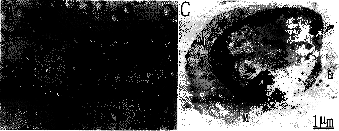
[0073] Freshly isolated cells were fixed with 2.5wt% cold glutaraldehyde for 30min, washed twice with PBS, fixed with 1wt% cold starvation acid for 4 hours, and then rinsed with PBS for 3 times; dehydrated with different concentrations of methanol, embedded in resin, ultrathin The sections were stained with uranyl acetate and lead citrate, and the ultrastructure of the cells was observed under a Philips EM 301 electron microscope ( image 3 ).
[0074] Observing the oval cells obtained in Example 3 under an electron microscope, it was found that the ultrastructural characteristics of the isolated cells were: ① oval nucleus; ② less cytopl...
PUM
 Abstract
Description
Claims
Application Information
 - R&D
- Intellectual Property
- Life Sciences
- Materials
- Tech Scout
- Unparalleled Data Quality
- Higher Quality Content
- 60% Fewer Hallucinations
Browse by: Latest US Patents, China's latest patents, Technical Efficacy Thesaurus, Application Domain, Technology Topic, Popular Technical Reports.
© 2025 PatSnap. All rights reserved.Legal|Privacy policy|Modern Slavery Act Transparency Statement|Sitemap|About US| Contact US: help@patsnap.com


