Medical image-based intraoperative auxiliary display method and device and storage medium
An auxiliary display and medical imaging technology, applied in the field of CDN, can solve the problems of inability to practically apply surgery, inconvenient understanding, and inability to determine the planning of the surgical path, so as to achieve the effect of improving the success rate of surgery, improving accuracy, and improving auxiliary efficiency
- Summary
- Abstract
- Description
- Claims
- Application Information
AI Technical Summary
Problems solved by technology
Method used
Image
Examples
Embodiment 1
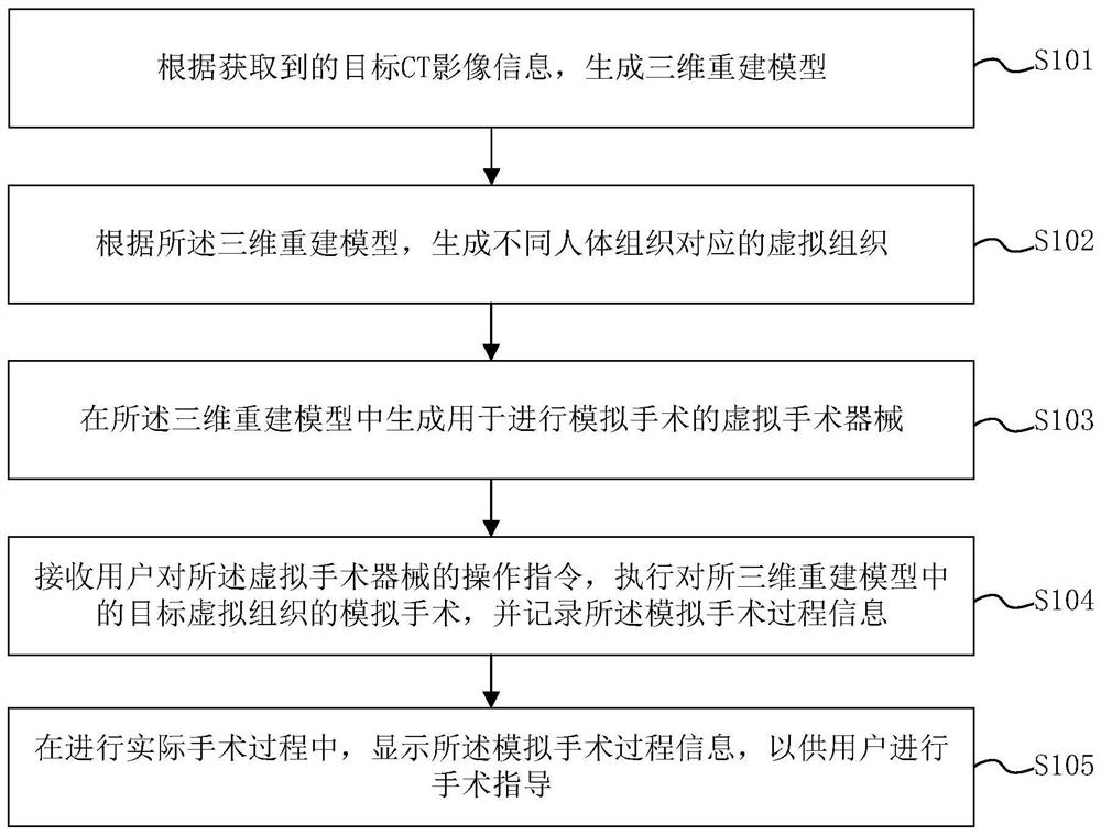
[0055] see figure 1 As shown, the embodiment of the present invention provides an intraoperative auxiliary display method based on medical images, and the method includes the following steps:
[0056] Step S101, generating a three-dimensional reconstruction model according to the acquired target CT image information;
[0057] Step S102, generating virtual tissues corresponding to different human tissues according to the three-dimensional reconstruction model;
[0058] Step S103, generating virtual surgical instruments for performing simulated surgery in the three-dimensional reconstruction model;
[0059] Step S104, receiving the user's operation instruction on the virtual surgical instrument, performing a simulated operation on the target virtual tissue in the three-dimensional reconstruction model, and recording the simulated operation process information;
[0060] Step S105 , during the actual operation process, display the simulated operation process information for the ...
Embodiment 2
[0095] see Figure 4 As shown, the embodiment of the present invention provides an intraoperative auxiliary display device based on medical images, and the device includes the following modules:
[0096] The first generation module 41 is used to generate a three-dimensional reconstruction model according to the acquired target CT image information;
[0097] The second generating module 42 is configured to generate virtual tissues corresponding to different human tissues according to the three-dimensional reconstruction model;
[0098] The third generating module 43 is used to generate a virtual surgical instrument for performing a simulated operation in the three-dimensional reconstruction model;
[0099] The recording module 44 is configured to receive the user's operation instruction for the virtual surgical instrument, perform a simulated operation on the target virtual tissue in the three-dimensional reconstruction model, and record the simulated operation process informa...
Embodiment 3
[0104] The embodiment of the present invention also provides a computer device, which may be a computing device such as a desktop computer, a notebook computer, a palmtop computer, or a cloud server. like Figure 5 As shown, the device may include, but is not limited to, a processor and a memory, wherein the processor and the memory may be connected through a bus or in other ways.
[0105] The processor can be a central processing unit (Central Processing Unit, CPU) or other general-purpose processors, digital signal processors (Digital Signal Processor, DSP), graphics processing units (Graphics Processing Unit, GPU), embedded neural network processors (Neural-network Processing Unit, NPU) or other dedicated deep learning coprocessor, Application Specific Integrated Circuit (ASIC), Field-Programmable Gate Array (Field-Programmable Gate Array, FPGA) or other programmable logic devices , discrete gate or transistor logic devices, discrete hardware components and other chips, or...
PUM
 Abstract
Description
Claims
Application Information
 - R&D Engineer
- R&D Manager
- IP Professional
- Industry Leading Data Capabilities
- Powerful AI technology
- Patent DNA Extraction
Browse by: Latest US Patents, China's latest patents, Technical Efficacy Thesaurus, Application Domain, Technology Topic, Popular Technical Reports.
© 2024 PatSnap. All rights reserved.Legal|Privacy policy|Modern Slavery Act Transparency Statement|Sitemap|About US| Contact US: help@patsnap.com


