Image denoising method, electronic equipment and storage medium
An image and memory technology, applied in the field of medical image processing, can solve problems such as the inability to guarantee the consistency of scanned content information, achieve the effect of reducing noise levels and meeting consistency requirements
- Summary
- Abstract
- Description
- Claims
- Application Information
AI Technical Summary
Problems solved by technology
Method used
Image

Examples
Embodiment 1
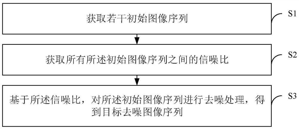
[0042] This embodiment provides an image denoising method for performing denoising processing on an image sequence of perfusion images. Such as figure 1 As shown, the method specifically includes the following steps:
[0043] S1. Acquire several initial image sequences.
[0044] In this embodiment, the several initial image sequences refer to the image sequences of perfusion images, for example, the perfusion images may be CTP images X scanned by CT equipment, where X∈(T×P×W×H), where T represents The number of CT images collected during the CT scanning process, P, W, and H respectively represent the number of layers, width and height of the CT images collected at each moment. Wherein, according to the tissues and organs of the perfusion imaging, the CTP images may include brain CTP images, liver CTP images, cardiac CTP images, and the like.
[0045] S2. Obtain signal-to-noise ratios among all the initial image sequences.
[0046] In this embodiment, the signal-to-noise ra...
Embodiment 2
[0093] Such as Figure 4As shown, the present invention provides an image denoising system, including: a sequence acquisition module 11 , a signal-to-noise ratio acquisition module 12 and a denoising module 13 . Wherein, the sequence acquisition module 11 is used to acquire several initial image sequences; the signal-to-noise ratio acquisition module 12 is used to acquire the signal-to-noise ratio between all the initial image sequences; the denoising module 13 is used to, based on the signal-to-noise ratio, The initial image sequence is subjected to denoising processing to obtain a target denoising image sequence.
[0094] Preferably, the denoising module 13 is specifically used for:
[0095] performing denoising processing on the initial image sequence to obtain an initial denoising image sequence;
[0096] Taking the initial denoised image sequence as a new initial image sequence, and returning to the step of obtaining the signal-to-noise ratio between the initial image s...
Embodiment 3
[0112] This embodiment provides an electronic device, which can be expressed in the form of a computing device (for example, it can be a server device), including a memory, a processor, and a computer program stored on the memory and operable on the processor, wherein the processor The image denoising method provided in Embodiment 1 can be realized when the computer program is executed.
[0113] Figure 5 A schematic diagram of the hardware structure of this embodiment is shown, as Figure 5 As shown, the electronic device 9 specifically includes:
[0114] At least one processor 91, at least one memory 92, and a bus 93 for connecting different system components, including the processor 91 and the memory 92, wherein:
[0115] The bus 93 includes a data bus, an address bus, and a control bus.
[0116] The memory 92 includes a volatile memory, such as a random access memory (RAM) 921 and / or a cache memory 922 , and may further include a read only memory (ROM) 923 .
[0117] M...
PUM
 Abstract
Description
Claims
Application Information
 - R&D
- Intellectual Property
- Life Sciences
- Materials
- Tech Scout
- Unparalleled Data Quality
- Higher Quality Content
- 60% Fewer Hallucinations
Browse by: Latest US Patents, China's latest patents, Technical Efficacy Thesaurus, Application Domain, Technology Topic, Popular Technical Reports.
© 2025 PatSnap. All rights reserved.Legal|Privacy policy|Modern Slavery Act Transparency Statement|Sitemap|About US| Contact US: help@patsnap.com