
Method for modeling mouse far-end optic nerve injury model through skull positioning puncture

An optic nerve injury and mouse technology, applied in medical science, veterinary instruments, veterinary surgery, etc., can solve the problems of modeling failure, distal optic nerve injury, high mortality, etc., and achieve a simple and easy modeling method. Effect

Pending Publication Date: 2021-12-07
RUIJIN HOSPITAL AFFILIATED TO SHANGHAI JIAO TONG UNIV SCHOOL OF MEDICINE
View PDF0 Cites 0 Cited by
  • Summary
  • Abstract
  • Description
  • Claims
  • Application Information

AI Technical Summary

Problems solved by technology

This method quantifies the force transmitted to the ocular surface, but brow blasts cause severe anterior-posterior segment injury with high mortality rates (ranging from 24% to 46% depending on the level of air pressure applied). Wait)
[0009] (4) The SI-TON model is to place the microtip probe ultrasonic instrument on the orbit just above the entrance of the optic nerve into the bone canal to transmit ultrasonic pulses, but the sound velocity difference between air and bone makes it difficult for sound waves to focus in such a narrow space , which can easily lead to modeling failure
[0010] The above methods can simulate optic nerve injury to a certain extent, but each has its own defects, and none of them involves distal optic nerve injury. Therefore, it is necessary to develop a stable, effective and simple mouse distal optic nerve injury model to carry Relevant basic research in the field

Method used


Image

  • Method for modeling mouse far-end optic nerve injury model through skull positioning puncture
  • Method for modeling mouse far-end optic nerve injury model through skull positioning puncture
  • Method for modeling mouse far-end optic nerve injury model through skull positioning puncture

Examples


Embodiment 1

[0035] The construction method of a mouse distal optic nerve injury model provided in this embodiment is as follows:

[0036] (1) Modeling environment: room temperature 22-24 ℃, well ventilated;

[0037] (2) Anesthesia: male 4-6 week BALA / C mice were injected intraperitoneally with 10 mg / kg of xylazine and 25 mg / kg of ketamine hydrochloride to anesthetize the mice;

[0038] (3) Skin preparation positioning: remove the hair on the top of the head, cut the skin on the top of the head, and bluntly separate the fascia to expose the anterior fontanelle. Use a cranial stereotaxic instrument to find the body surface position corresponding to the optic nerve. Starting from the anterior fontanel, move to the tail and right side respectively. Move 0.5mm as the needle entry point;

[0039] (4) Needle insertion: The 27G needle was inserted slowly. When the needle was inserted to about 3mm, a breakthrough was felt and the right eyeball fluttered. This was a reaction to the optic nerve inj...

experiment example 1

[0042] 1. The modeling effect of the ONT model constructed by skull puncture

[0043] 1) Survival rate: During the observation period of 4 weeks, among 180 mice modeled with ONT, 154 survived, and the survival rate was 85.56%.

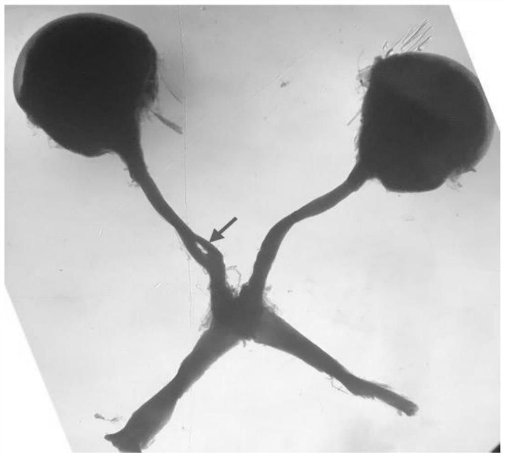
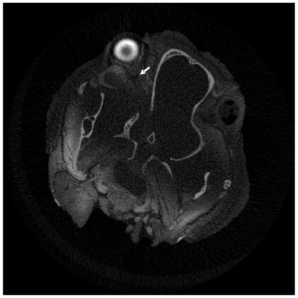
[0044] 2) Success rate: 1 week after modeling, 20 mice were subjected to optic nerve separation and exposure, and observed under a microscope after fixation, 18 of them showed damage to the right distal optic nerve ( figure 1 ), the modeling success rate is estimated to be 90%. Another 20 mice were observed with microCT for optic nerve damage, and 19 of them had optic nerve damage ( figure 2 ), the modeling success rate is estimated to be 95%. Combining the two evaluation methods, the estimated modeling success rate is about 92.5%.

[0045] 2. Brn3a staining of retinal slices to observe the survival of retinal ganglion cells

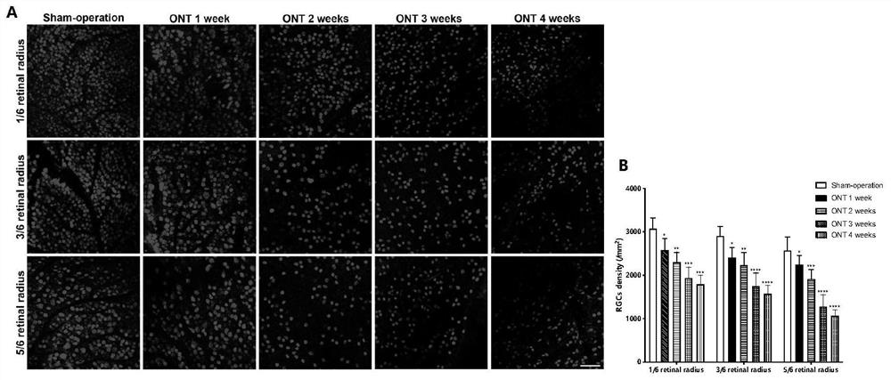
[0046] 1, 2, 3, and 4 weeks after ONT modeling, retinal spread Brn3a staining showed that the density of RGCs in the 1 / 6, ...


PUM


Abstract

The invention provides a method for modeling a mouse far-end optic nerve injury model through skull positioning puncture and belongs to the technical field of clinical animal models. The modeling method comprises the following steps: (1) anesthetization: injecting an anesthetic into an abdominal cavity of a mouse; (2) preserved skin positioning: removing top hair from the mouse, cutting open top skin, bluntly separating a fascia, and exposing an anterior fontanelle, wherein a body surface position corresponding to an optic nerve is found through a head stereotaxic instrument, the anterior fontanelle serves as a starting point, and a position moving 0.5mm towards the tail and the right side separately from the anterior fontanelle serves as a needle inserting point; and (3) needle insertion: slowly inserting a 27G needle to reach a skull base when the needle insertion depth of the 27G needle is 6mm, then, slowly pulling out the needle, carrying out pressing to stop bleeding after pulling out the needle, and suturing the skull top fascia and the skin, so as to prevent infection after operation. According to the model, the survival rate of injured mice can reach 85.56%, the modeling success rate for optic nerve injury can reach 95%, and especially the modeling success rate for far-end optic nerve injury can reach 90%.

Description

technical field [0001] The invention belongs to the technical field of clinical animal models, and in particular relates to a method for establishing a mouse distal optic nerve injury model by cephalometric puncture. Background technique [0002] Traumatic optic neuropathy (TON) is a serious complication of craniocerebral, orbital and facial trauma. Post-injury visual impairment is severe and often leaves permanent visual impairment. Clinically, the incidence of TON has been reported to be 0.5% in closed craniocerebral injuries and 2.5% in maxillofacial injuries, with motor vehicle and bicycle accidents accounting for the majority, followed by falls and assaults. Optic nerve injury can be divided into two types: direct injury and indirect injury. Direct optic nerve injury refers to orbital trauma that penetrates the interior or exterior of the fluoroscopy neural canal, resulting from direct destruction of the optic nerve by bone fragments or hematomas; indirect optic nerve ...

Claims


Application Information

Patent Timeline
Patent Type & Authority Applications(China)
IPC IPC(8): A61D1/00A01K67/02
CPCA61D1/00A01K67/02A01K2227/105A01K2267/03
Inventor 钟一声余欢沈柄桥钟慧敏章敏贵陈珺珏
Owner RUIJIN HOSPITAL AFFILIATED TO SHANGHAI JIAO TONG UNIV SCHOOL OF MEDICINE
Learn More