
Pathological tissue section staining kit

A tissue section and staining reagent technology, applied in the field of pathological tissue section staining, can solve the problems of increasing the control difficulty of differentiation, indistinct coloring of cell nuclei, and great harm to the operator, achieving clear coloring effect, increasing staining ability, good effect

Pending Publication Date: 2021-12-03
SHANDONG JUNTENG MEDICAL TECH CO LTD
View PDF0 Cites 2 Cited by
  • Summary
  • Abstract
  • Description
  • Claims
  • Application Information

AI Technical Summary

Problems solved by technology

[0003] In the conventional H&E staining process, xylene is used for dewaxing and subsequent transparent mounting, which is very harmful to the operator; the hydration and dehydration process uses volatile and flammable chemicals such as graded alcohol; the differentiation solution usually uses hydrochloric acid Alcohol differentiation, the use of hydrochloric acid on the one hand increases the difficulty of differentiation control, on the other hand, tissue slices with poor pretreatment are prone to chipping during the differentiation process, which affects diagnosis and causes cross-contamination; ammonia solution is often used in the blue-return solution , the stock solution of ammonia water is slightly different in concentration due to different storage and manufacturers, which causes the concentration difference of the prepared bluing liquid, which is easy to cause batch-to-batch difference; For conversion, chemical oxidants are often used, such as mercury oxide, hydrogen peroxide and sodium iodate, but the use of chemical oxidants usually produces ineffective reaction products such as oxidized hematin and complex polymer precipitation. The hematoxylin staining solution prepared by this method is more than The naturally mature Ehrlich'shematoxylin (Ehrlich'shematoxylin) fails faster, resulting in a short validity period of the stain solution, poor coloring effect, and unclear coloring of the nucleus

Method used


Image

  • Pathological tissue section staining kit
  • Pathological tissue section staining kit
  • Pathological tissue section staining kit

Examples


Embodiment 1

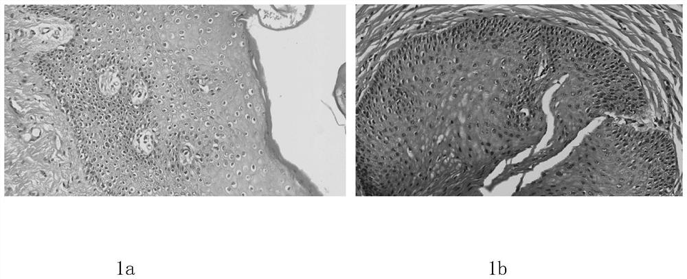
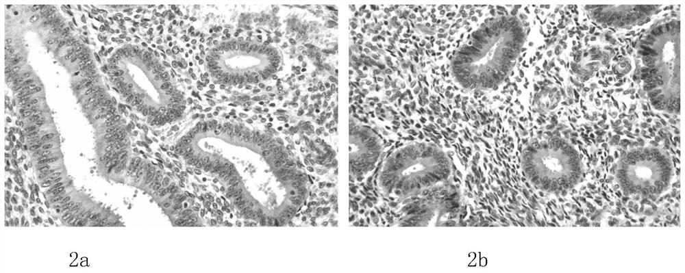
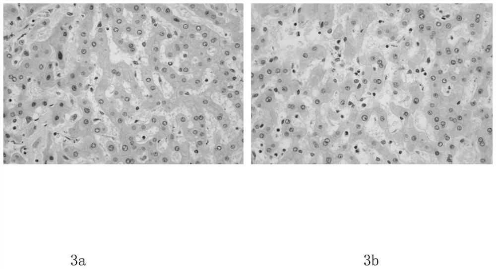
[0037] 1. Experimental objects: pathological tissues (perianal, uterus, liver, kidney, intestine and breast), all pathological tissues are taken from human isolated diseased tissues.

[0038] 2. Staining reagents: pathological tissue section staining kit / conventional H&E staining reagents.

[0039] 3. Experimental process:

[0040] (1) Weigh the required raw materials for section dewaxing solution, conversion solution, cleaning solution, differentiation solution, bluing solution, hematoxylin staining solution, and eosin staining solution according to the following proportions;

[0041] The components in parts by mass and volume of the slice dewaxing liquid are: 80 parts of solvent oil D60, 20 parts of n-dodecane;

[0042] The mass and volume parts of the conversion liquid are divided into: 50 parts of dipropylene glycol methyl ether and 50 parts of propylene glycol methyl ether acetate;

[0043]The components of the mass volume fraction of the cleaning solution are: 70 parts...


PUM


Abstract

The invention relates to a pathological tissue section staining kit, which belongs to the field of pathological tissue section staining and comprises a section dewaxing solution, a conversion solution, a cleaning solution, a differentiation solution, a blue returning solution, a hematoxylin staining solution, an eosin staining solution and a mounting medium, the invention provides the novel pathological tissue section staining kit which is simple in component, good in effect and low in toxicity, and the preparation method, and the kit is mainly used for pathological tissue H&E staining. The method is characterized in that 1, the harm of traditional dyeing reagents such as dimethylbenzene and alcohol to an operator is reduced; 2, the pollution to the environment is reduced; 3, extra deionized water does not need to be introduced in the dyeing process; 4, a differentiation solution and a blue returning solution are designed by adopting a buffer system, so that the effect is milder, and the coloring effect of H&E dyeing is clearer and brighter; and 5, compared with a traditional hematoxylin formula, beta-cyclodextrin is introduced into the kit to neutralize a product iodine reduced by sodium iodate, so that the dyeing capability of the staining solution is improved, and meanwhile, a reducing agent dibutylated hydroxytoluene is introduced, so that the service life and the shelf life of the staining solution are prolonged.

Description

technical field [0001] The invention belongs to the field of pathological tissue section staining, in particular to a pathological tissue section staining kit. Background technique [0002] In life sciences such as histopathology and microbiology, specimens are often examined using a microscope (such as a light microscope), which requires the specimen to be mounted on a glass slide, stained, and then stained using a glass coverslip with an adhesive. cover film. Hematoxylin and eosin staining (H&E) is commonly used to stain tissue samples during pathological examination. H&E staining is the complex and tedious steps of dewaxing, hydration, hematoxylin staining, water washing, differentiation, water washing, bluing, water washing, eosin staining, dehydration, transparent and mounting of tissue sections, so that the tissue cells Different components are dyed in different shades of color to enhance the contrast between different components, making it easier for pathologists to...

Claims


Application Information

Patent Timeline
IPC IPC(8): G01N1/30G01N1/31
CPCG01N1/30G01N1/31G01N2001/302
Inventor 郭玉川孙玉芬吴业冰王亮夏国建孙丽莹王文杰
Owner SHANDONG JUNTENG MEDICAL TECH CO LTD
Learn More