
Micro-fluidic cell chip and virus isolated culture method based on same

A cell chip and virus isolation technology, applied in the field of biological sciences, can solve the problem of rapid isolation and culture of virus-sensitive host cells, high-throughput screening of viruses, difficulty in obtaining infected samples of major viral infectious diseases, virus classification, and identification and vaccine development obstacles, etc., to achieve the effect of improving sample utilization, increasing throughput processing capacity, and shortening separation and identification time

Pending Publication Date: 2021-11-12
WUHAN INST OF VIROLOGY CHINESE ACADEMY OF SCI
View PDF0 Cites 2 Cited by
  • Summary
  • Abstract
  • Description
  • Claims
  • Application Information

AI Technical Summary

Problems solved by technology

This traditional culture method usually requires a large number of cells (10 5 -10 8 1), the use of medium and other reagents is large, the experimental period is long (7-10 days), and the sensitivity is low; for major viral infectious diseases, the clinical samples are often more precious, when the viral load in the sample is low, The possibility of failure of isolation and culture is extremely high; if the isolated and cultured virus lacks an in vitro culture system (such as human norovirus), it is impossible to develop an effective antiviral strategy
At present, when the virus is isolated and identified through cell culture and infection, it is mainly to inoculate different cell lines in separate cell culture flasks or in the same microplate and then infect them separately. The operation steps are cumbersome and prone to cross-contamination. Realize rapid and high-throughput screening of virus-sensitive host cells and rapid isolation and culture of viruses
[0004] In actual work, it is difficult to obtain infected samples of major viral infectious diseases. Even if the virus-containing material is obtained, the viral load is very low after treatment, and it is almost impossible to isolate and culture by conventional methods. Virus taxonomy, identification, and vaccine development pose major obstacles and waste precious virus samples

Method used


Image

  • Micro-fluidic cell chip and virus isolated culture method based on same
  • Micro-fluidic cell chip and virus isolated culture method based on same
  • Micro-fluidic cell chip and virus isolated culture method based on same

Examples


Embodiment 1

[0052] Example 1, Simulated screening of sensitive host cells of enterovirus EV71 (Chinese vaccine strain, GenBank No. HQ328793) in a microfluidic cell chip at a low multiplicity of infection (MOI).

[0053] The cell line can be a conventional virus-amplified cell line, or an in vitro cultured cell line from other species.

[0054] (1) Characterization of typical CPE (cytopathic effect) of EV71: human lung cancer cells (A549), African green monkey kidney cells (Vero cells) and human malignant embryonic rhabdoid tumor cells (RD cells), 1×10 4 The number of cells was seeded into a 96-well plate at 37°C, 5% CO 2 After culturing in the incubator for 12 hours, EV71 was infected at MOI=40.0; 24 hours and 36 hours after infection, the cytopathic changes were observed with a bright field inverted microscope. The results showed that all three types of cells produced cytopathic changes, decreased cell adhesion ability, rounded and shrunken cell shape, and poor light transmission, among...

Embodiment 2

[0056] Example 2, Simulated screening of EV71 sensitive host cells in a microfluidic cell chip at extremely low MOI.

[0057] The cell line can be a conventional virus-amplified cell line, or an in vitro cultured cell line from other species.

[0058] Very low MOI infection: A549, Vero and RD cells at 1 x 10 4 The number of cells was seeded into the microfluidic cell chip culture chamber at 37°C, 5% CO 2 Cultured in the incubator for 12h, EV71 at MOI=3.89×10 -4 Infection was carried out; cytopathic changes were observed with a bright-field inverted microscope 48 hours after infection. The results show that RD cells produce cytopathy the earliest and are the most significant, consistent with the results in the 96-well plate in Example 1 (such as Image 6 shown).

Embodiment 3

[0059] Example 3, Simulated screening of influenza virus (A / Puerto Rico / 8 / 1934, H1N1) sensitive host cells in a microfluidic cell chip at high MOI.

[0060] The cell line can be a conventional virus-amplified cell line, or an in vitro cultured cell line from other species.

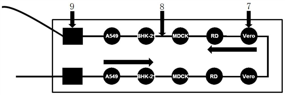
[0061] (1) H1N1 typical CPE characterization: A549, baby hamster kidney cells (BHK-21), canine kidney cells (MDCK), Vero and RD cells, 1×10 4 The number of cells was seeded into a 96-well plate at 37°C, 5% CO 2 After being cultured in an incubator for 12 hours, H1N1 was infected at MOI=12.3; 24 hours after infection, cytopathic changes were observed with a bright-field inverted microscope. The results showed that MDCK cells produced obvious cytopathic changes, the inherent shape of the cells disappeared, the cells fell off and produced vacuoles, and other cell lines had no obvious lesions (such as Figure 7 shown).

[0062] (2) High MOI infection: A549, BHK-21, MDCK, Vero and RD cells, at 1×10 4 The nu...


PUM


Abstract

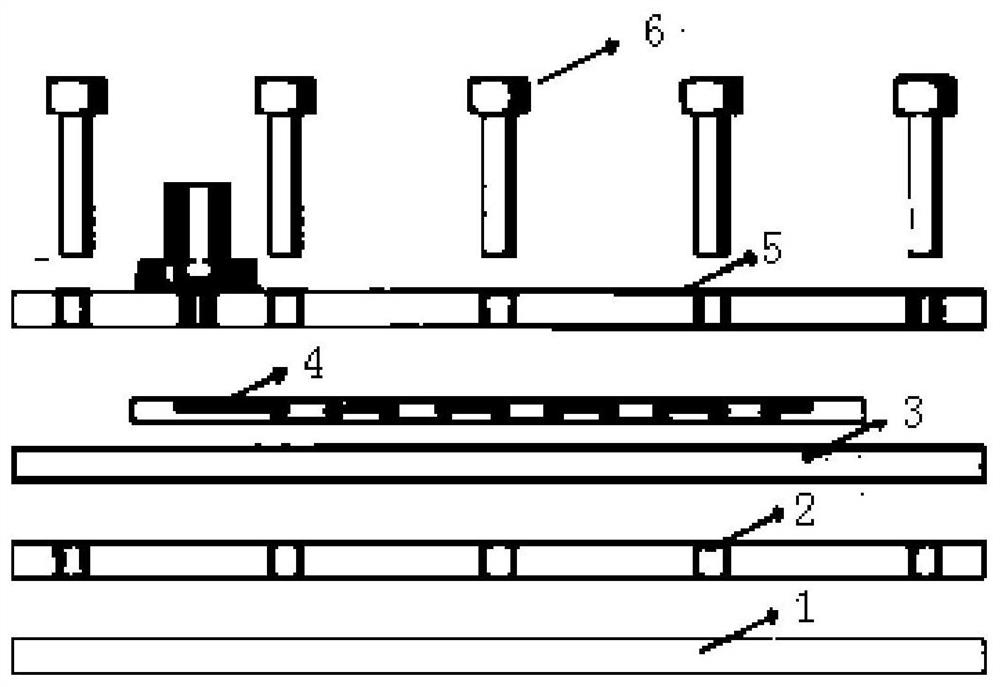
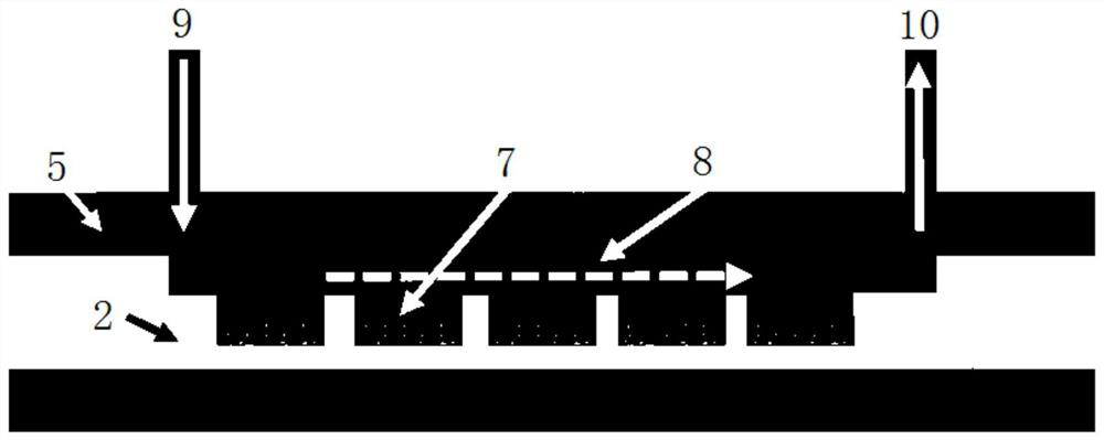
The invention discloses a micro-fluidic cell chip and a virus isolated culture method based on same. The micro-fluidic cell chip comprises a metal frame, a lower cover plate, a chip frame, a core chip and an upper cover plate, wherein a plurality of cell culture chambers are formed in the core chip; a main runner is formed in the surface of the core chip; and the main runner is communicated with the plurality of cell culture chambers. According to the virus isolated culture method based on the micro-fluidic cell chip, a method of co-culturing multiple cell lines is adopted, a to-be-analyzed sample and different cell lines are sequentially incubated, clinical samples can be obviously saved, and the sample utilization rate is improved to the greatest degree. Meanwhile, the consumption of a culture reagent can be reduced, and the separation and identification time is shortened. Because the micro-fluidic cell chip provided by the invention contains the plurality of built-in cell culture chambers, culture of at least ten different cell lines can be simultaneously realized theoretically, the screening flux of virus sensitive host cells is obviously increased, and high-flux, automatic and rapid screening of the virus sensitive cell lines is realized.

Description

technical field [0001] The invention belongs to the field of biological sciences, and in particular relates to a microfluidic cell chip and a virus isolation and culture method based on the cell chip. Background technique [0002] At present, new and re-emerged viral infectious diseases still pose a serious threat to human life and property. Therefore, the rapid identification of viruses is crucial to the early diagnosis of diseases and the prevention and control of epidemics. As we all know, the virus plaque test based on living cell culture is the gold standard for virus diagnosis and follows the "Koch's law". When a new viral infectious disease occurs, an important issue is to identify the sensitive host cell line allowed by the virus by plaque test, which is especially critical for subsequent virus amplification, virus isolation and culture, and vaccine production. [0003] Existing conventional virus isolation and culture methods are usually carried out in polystyrene...

Claims


Application Information

Patent Timeline
IPC IPC(8): C12M3/00C12N7/00C12R1/93
CPCC12M23/16C12M23/46C12N7/00B01L3/502761B01L3/5027C12N2770/32352C12N2760/16152
Inventor 李峰贺永苏炜德邱京江
Owner WUHAN INST OF VIROLOGY CHINESE ACADEMY OF SCI
Learn More