3D printing implant as well as preparation method and application thereof

A 3D printing and 3D printer technology, applied in the field of bionics, can solve the problems of reducing inflammation, low mechanical strength of tissue regeneration membrane, low dimensional accuracy, etc., achieving good biocompatibility, high mechanical and mechanical properties, dimensional stability, and inflammatory response. low effect

Active Publication Date: 2021-09-21
BEIJING TONGREN HOSPITAL AFFILIATED TO CAPITAL MEDICAL UNIV
View PDF7 Cites 0 Cited by
  • Summary
  • Abstract
  • Description
  • Claims
  • Application Information

AI Technical Summary

Problems solved by technology

[0007] The purpose of the present invention is to overcome the problems of low mechanical strength and low dimensional accuracy of the guiding tissue regeneration membrane existing in the prior art, and provide a 3D printing implant and its preparation method and application. The 3D printing implant has high The dimensional accuracy is used in the anti-scar after glaucoma filtration surgery. It can inhibit the scarring area of ​​the filtering bleb and can inhibit the activity of fibroblasts to realize the function of inhibiting the healing of the scarring of the filtering bleb; its promoting healing area can promote the activity of fibroblasts To achieve the function of promoting the healing of the conjunctival flaps on both sides; at the same time, it can reduce inflammation and have sufficient support to resist the contraction of surrounding tissues so as to maintain intraocular pressure and filter bleb shape for a long time

Method used


Image

  • 3D printing implant as well as preparation method and application thereof
  • 3D printing implant as well as preparation method and application thereof
  • 3D printing implant as well as preparation method and application thereof

Examples


preparation example Construction

[0031] The second aspect of the present invention provides a method for preparing the above-mentioned 3D printed implant, wherein the method includes:

[0032] The photocurable and degradable material is printed layer by layer by using a photocurable 3D printer to obtain the 3D printed implant.

[0033] In the present invention, the above-mentioned implants are prepared using micro-nano-scale 3D printing technology.

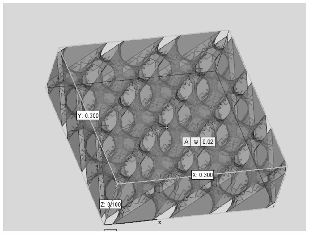
[0034] In a specific embodiment of the present invention, the preparation method of the 3D printing implant includes: using surface projection micro stereolithography (Projection Micro Litho Stereo Exposure, PuLSE) technology to make the implant, specifically: using high-precision ultraviolet The photolithographic projection system projects the pattern to be printed onto the liquid surface of the resin tank, and solidifies the resin on the liquid surface. The rapid micro-stereoscopic molding of the implant can be realized through the precise movement of the sampl...

Embodiment

[0066] according to figure 1 Print the drawings shown and prepare the 3D printed implants as follows: The photocurable degradable material was printed using the nanoArch P130, and the printer's own camera was used to image each layer during processing. To remove residual photoinitiator and thermal curing agent, the membrane was carefully washed by immersing it in a large volume of deionized water (500 mL) for 1.5 hours. Replace the water with fresh water every 0.5 hours. The photocurable and degradable material includes 100 parts of polylactic acid-acrylate, 1.5 parts of photoinitiator 1173 and 1.5 parts of trimethylhexamethylenediamine. The light source power of the 3D printer is 20mW / cm 2 , the thickness of each layer is 0.02mm, and the printing time of each layer is 6s.

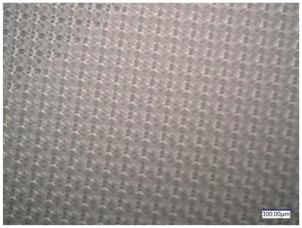
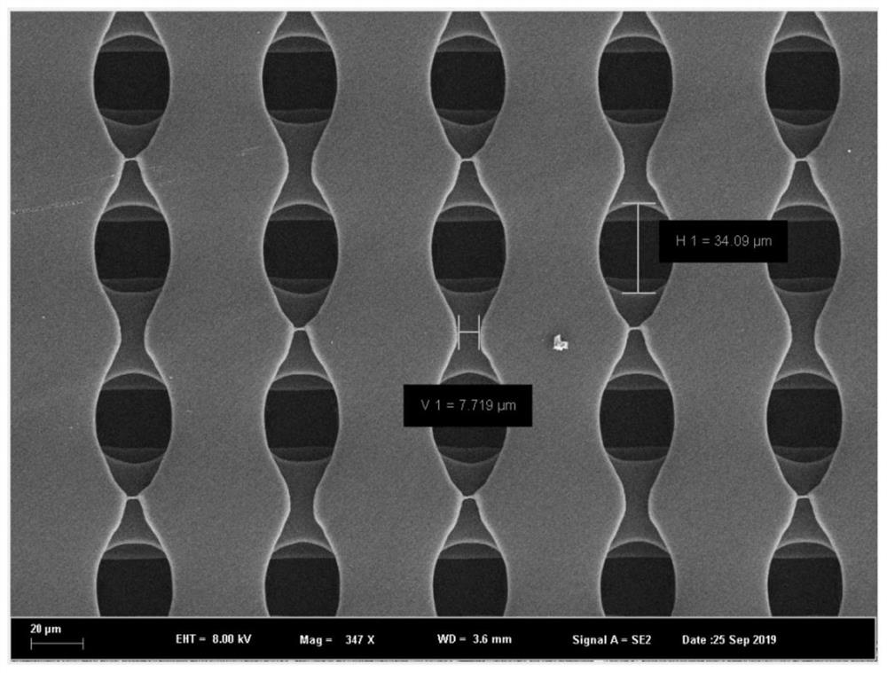
[0067] The morphology and physical and mechanical properties of the prepared 3D printed implants were tested, and the results are shown in Table 1.

[0068] figure 2 Shown is the micrograph of the 3D...


PUM

PropertyMeasurementUnit
diameteraaaaaaaaaa
diameteraaaaaaaaaa
thicknessaaaaaaaaaa

Abstract

The invention relates to the field of biological materials, in particular to a 3D printing implant and a preparation method and application thereof. The implant comprises a plurality of three-dimensional porous structure layers, each three-dimensional porous structure layer is provided with a plurality of convex bionic microstructures, and pores exist between the bionic microstructures; and pores in the three-dimensional porous structure layers are communicated with one another. The 3D printed implant has high dimensional precision and is used for resisting scars after glaucoma filtration, and a filtration vesicle scar inhibition area of the 3D printed implant can inhibit the activity of fiber cells so as to realize the healing function of inhibiting filtration vesicle cicatrization; the healing promoting area can promote the activity of fibroblasts so as to realize the function of promoting the healing of conjunctival flaps on the two sides; meanwhile, inflammation can be reduced, and sufficient supporting force is achieved to resist shrinkage of surrounding tissue, so that intraocular pressure and the form of filtered bubbles are maintained permanently.

Description

technical field [0001] The invention relates to the field of bionics, in particular to a 3D printing implant and its preparation method and application. Background technique [0002] Glaucoma filtering surgery (GFS) is the most widely used anti-glaucoma surgery, which reduces intraocular pressure by creating a new aqueous humor outflow channel between the sclera and subconjunctiva. The success of the operation needs to achieve a delicate balance between maintaining the watertightness of the filtering bleb and preventing the formation of scarring in the filtering area. Healing; ②Promote the healing of the conjunctival flap and prevent the filtering bleb from communicating directly with the outside world. However, the conjunctival flap, scleral bed, scleral flap, and Tenon's capsule are closely adjacent without other tissue separation. How to meet the above-mentioned different healing requirements at the same time is the key to ensuring long-term intraocular pressure control ...

Claims


Application Information

Patent Timeline
Patent Type & Authority Applications(China)
IPC IPC(8): A61L31/14A61F9/007B33Y80/00
CPCA61L31/148A61L31/146B33Y80/00A61F9/00781A61F2009/00891Y02P10/25
Inventor 甄毅毛迎燕王宁利
Owner BEIJING TONGREN HOSPITAL AFFILIATED TO CAPITAL MEDICAL UNIV
Why Eureka
Try Eureka