BFGF mesenchymal stem cell exosomes and preparation method and application thereof
A technology of mesenchymal stem cells and exosomes, applied in the field of bFGF mesenchymal stem cell exosomes and its preparation, can solve the problems of low specificity, low bFGF content, and inability to repair tissues well
- Summary
- Abstract
- Description
- Claims
- Application Information
AI Technical Summary
Problems solved by technology
Method used
Image


Examples
Embodiment 1
[0048] The construction of embodiment 1 lentiviral vector
[0049] 1. Design the sequence of the bFGF fusion protein, use the signal peptide and transmembrane region of the mesenchymal stem cell surface marker CD44 as the signal peptide and transmembrane region of the fusion protein, and select the flexible chain for the linker connecting the target protein and the transmembrane region. The signal peptide, target gene bFGF, linker, and the sequence of the transmembrane region are as follows: N-terminal signal peptide:
[0050] ATGGACAAGTTTTGGTGGCACGCAGCCTGGGGACTCTGCCTCGTGCCGCTGAGCCTGGCG (SEQ ID NO. 1)
[0051] The nucleotide sequence of the target protein bFGF:
[0052] CTGGTGGGTGTGGGGGGTGGAGATGTAGAAGATGTGACGCCGCGGCCCGGCGGGTGCCAGATTAGCGGACGCGGTGCCCGCGGTTGCAACGGGATCCCGGGCGCTGCAGCTTGGGAGGCGGCTCTCCCCAGGCGGCGTCCGCGGAGACACCCATCCGTGAACCCCAGGTCCCGGGCCGCCGGCTCGCCGCGCACCAGGGGCCGGCGGACAGAAGAGCGGCCGAGCGGCTCGAGGCTGGGGGACCGCGGGCGCGGCCGCGCGCTGCCGGGCGGGAGGCTGGGGGGCCGGGGCCGGGGCCGTGCCCCGGAGCG...
Embodiment 2
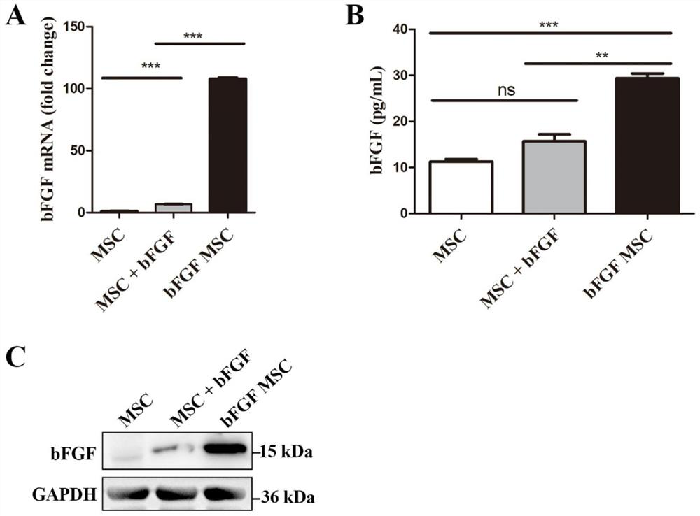
[0066] Example 2 Characterization of Mesenchymal Stem Cells Infected with Viruses
[0067]1. For lentivirus infection of mesenchymal stem cells, take 950 μL 1×HBS into the EP tube, add 10 μg bFGF plasmid, mix well, slowly add 50 μL CaCl2 solution (2M), mix well and let it stand for 20 minutes, then add to the cell state In 293T cells with a good density of about 60%-70%, mix gently, put in 37°C, 5% CO 2 After 12 hours, the medium was replaced with 10 mL of 30% FBS complete medium. After 48 hours, the cell supernatant was collected and centrifuged at 4000 rpm for 15 minutes at room temperature. Take the supernatant and add it to the mesenchymal stem cells that have grown to 50%-60%, add polybrene with a final concentration of 8ug / mL, mix well, and put it in 37°C, 5% CO 2 After 12 hours, replace with 10% FBS complete medium.
[0068] 2. Screening of bFGF mesenchymal stem cells, adding puromycin at a final concentration of 2ug / mL to the infected mesenchymal stem cells, and the ...
Embodiment 3
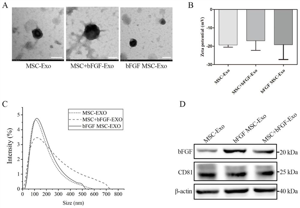
[0081] Example 3 Characterization of exosomes derived from mesenchymal stem cells integrating bFGF fusion protein gene
[0082] 1. Extract wild-type mesenchymal stem cells, mesenchymal stem cells incubated with bFGF growth factor and exosomes of mesenchymal stem cells integrating bFGF fusion protein gene (bFGF mesenchymal stem cell exosomes). Cultivate the three kinds of cells with 10% FBS complete medium. When the cell density reaches 70%, replace with 0.5% EV Free FBS medium. After 48h, harvest the cell culture supernatant, centrifuge at 500×g for 10min, and take the supernatant , centrifuge at 2000×g for 20min, then take the supernatant, centrifuge at 10000×g for 40min, take the supernatant, ultracentrifuge at 100000×g for 90min, take the precipitate, resuspend with appropriate amount of PBS, ultracentrifuge at 100000×g for 90min, collect the precipitate , resuspended in a small amount of PBS, and stored in an ultra-low temperature refrigerator. All the above centrifugatio...
PUM
 Abstract
Description
Claims
Application Information
 - R&D
- Intellectual Property
- Life Sciences
- Materials
- Tech Scout
- Unparalleled Data Quality
- Higher Quality Content
- 60% Fewer Hallucinations
Browse by: Latest US Patents, China's latest patents, Technical Efficacy Thesaurus, Application Domain, Technology Topic, Popular Technical Reports.
© 2025 PatSnap. All rights reserved.Legal|Privacy policy|Modern Slavery Act Transparency Statement|Sitemap|About US| Contact US: help@patsnap.com