Method for detecting exudate of retinal fundus image

A fundus image and detection method technology, applied in the field of medical detection, can solve the problems of inability to determine the patient's condition, the detection method is not precise enough, and the detection efficiency is low, so as to facilitate the diagnosis of the condition, save time, and improve the effect.

Pending Publication Date: 2021-03-02
黄珍珍
View PDF4 Cites 0 Cited by
  • Summary
  • Abstract
  • Description
  • Claims
  • Application Information

AI Technical Summary

Problems solved by technology

[0003] However, the existing technology is prone to low detection efficiency or insufficient detect

Method used


Image

  • Method for detecting exudate of retinal fundus image
  • Method for detecting exudate of retinal fundus image

Examples


Embodiment Construction

[0039] In order to make the object, technical solution and advantages of the present invention clearer, the present invention will be further described in detail below in conjunction with the accompanying drawings and embodiments. It should be understood that the specific embodiments described here are only used to explain the present invention, not to limit the present invention.

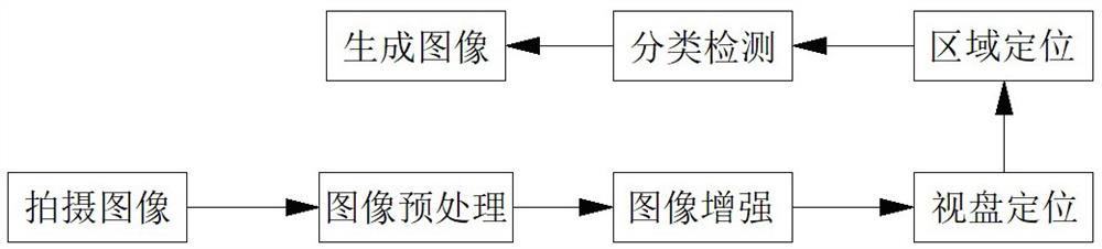
[0040] refer to Figure 1-2 , a kind of exudate detection method of retinal fundus image, the exudate detection method of retinal fundus image comprises the following steps:

[0041] Step 1: Take a fundus image with a non-mydriatic fundus camera;

[0042] Step 2: Preprocessing the fundus image;

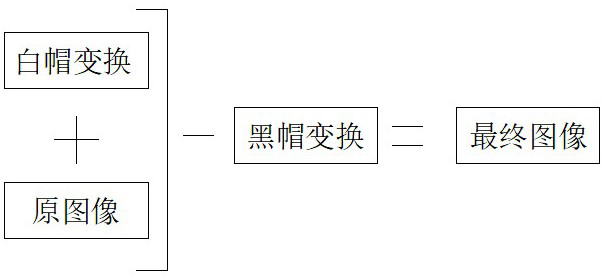
[0043] Step 3: Enhance the fundus image;

[0044] Step 4: Carry out optic disk positioning to the image;

[0045] Step 5: Locate the exudate area;

[0046] Step 6: Classify and detect the exudate area;

[0047] Step 7: Generate a fundus image of the marked exudate.

[0048] The non-mydriatic fundus ...


PUM


Abstract

The invention discloses a method for detecting exudate of a retinal fundus image. The method comprises the following steps: step 1, shooting a fundus image by using a mydriasis-free fundus camera; step 2, preprocessing the fundus image; step 3, enhancing the image; step 4, carrying out optic disk positioning on the image; step 5, positioning the exudate area; step 6, carrying out classification detection on the exudate areas; and step 7, generating a fundus image of the marked exudate. A color space model is used for preprocessing the fundus image, the image can be divided into color and grayscale information, the method is suitable for processing the gray scale image, the image enhancement effect is improved, the processed image is clearer, medical staff can judge the illness state conveniently, meanwhile, classification detection is conducted on the appearing area of suspicious penetrating fluid. After the detection is finished, the computer automatically prints a fundus image marked with the exudate.

Description

technical field [0001] The invention belongs to the technical field of medical detection, in particular to a method for detecting exudate of retinal fundus images. Background technique [0002] Retinal disease is a kind of eye disease, and its symptoms are mainly different degrees of visual impairment. For example, if the pyramidal cells in the macular area are damaged, central scotoma, central vision loss, and color vision disturbance may occur. Distortion or edema, and metamorphopsia may occur. In addition to a complete medical history review and routine eye exams, the ophthalmologist will do the following tests: Eye charts Retinopathy - Routine eye charts are used to measure the ability to see at different distances. Mydriasis examination - After dilating the pupil with eye drops, check the condition of the fundus and retina in detail. Intraocular pressure - a standard test to measure the pressure of fluid in the eyeball. Fundoscopy - The ophthalmologist does a detaile...

Claims


Application Information

Patent Timeline
IPC IPC(8): G06T7/00G06T5/30G06T5/00G06T5/40
CPCG06T7/0012G06T5/30G06T5/007G06T5/40G06T2207/10024G06T2207/30041G06T2207/20004
Inventor 黄珍珍
Owner 黄珍珍
Why Eureka
Try Eureka