
Auxiliary mammary duct in-situ cancer detection system

A breast duct and auxiliary detection technology, applied in the field of breast cancer detection, can solve the problems of low accuracy, insufficient computer image recognition accuracy, limiting the detection effect of breast duct carcinoma in situ, etc., so as to save workload and enhance accuracy. Effect

Active Publication Date: 2021-01-26
HENAN CANCER HOSPITAL
View PDF14 Cites 1 Cited by
  • Summary
  • Abstract
  • Description
  • Claims
  • Application Information

AI Technical Summary

Problems solved by technology

[0003] At present, the pathological biopsy of breast ductal cells mainly relies on pathologists to observe pathological tissue samples under a microscope, and make a diagnosis based on empirical knowledge such as cell morphology, which heavily relies on the experience of pathologists; in recent years, image recognition methods represented by deep learning have achieved For images with large heterogeneity such as breast cancer, the accuracy of image recognition is 70-80%, the accuracy of manual inspection of IDC by pathologists is less than 73.2%, and the accuracy of using CNN to identify IDC It is about 84%, and IDC and DCIS have high similarity in cell morphology, so it is difficult to distinguish only from cell morphology using CNN neural network, and the accuracy is not high
[0004] In the prior art, there are also some technical solutions for the auxiliary detection system of breast ductal carcinoma in situ. For example, a Chinese patent with application number 201710242892X discloses an auxiliary detection method for breast ductal carcinoma in situ: breast cancer Digital slices are manually marked to obtain images of DCIS and myoepithelial regions; read in digital slice file images, cut them into small pieces, and obtain whether the small piece of images contains DCIS or myoepithelial region images by querying the information in the file marked by the pathologist , so as to obtain three types of sample sets; start the neural network, establish a recognition model; identify the digital slices, find out the DCIS and myoepithelial regions, and record the corresponding probability; calculate the probability that each region is considered to be DCIS Probability; this technical solution uses the method of CNN to identify DCIS, and realizes DCIS automatic identification with high accuracy by simultaneously detecting DCIS cancer cells and myoepithelial tissue; however, the technical solution does not solve the problem that the accuracy of computer image recognition in the detection process is still insufficient problems, and the training of computer image recognition algorithms requires a large amount of sample data, which brings about the problem of preparing a large number of pathological sections, which limits the detection effect of breast ductal carcinoma in situ

Method used


Image

  • Auxiliary mammary duct in-situ cancer detection system
  • Auxiliary mammary duct in-situ cancer detection system
  • Auxiliary mammary duct in-situ cancer detection system

Examples


Embodiment approach

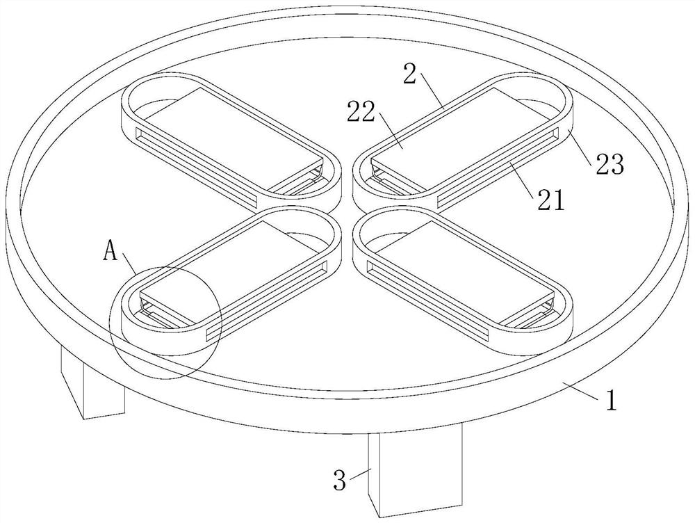
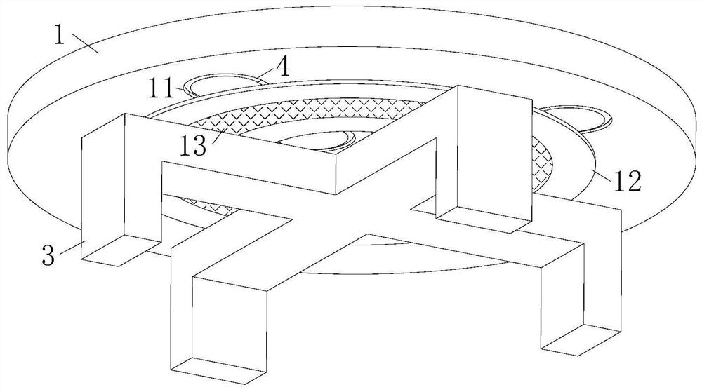
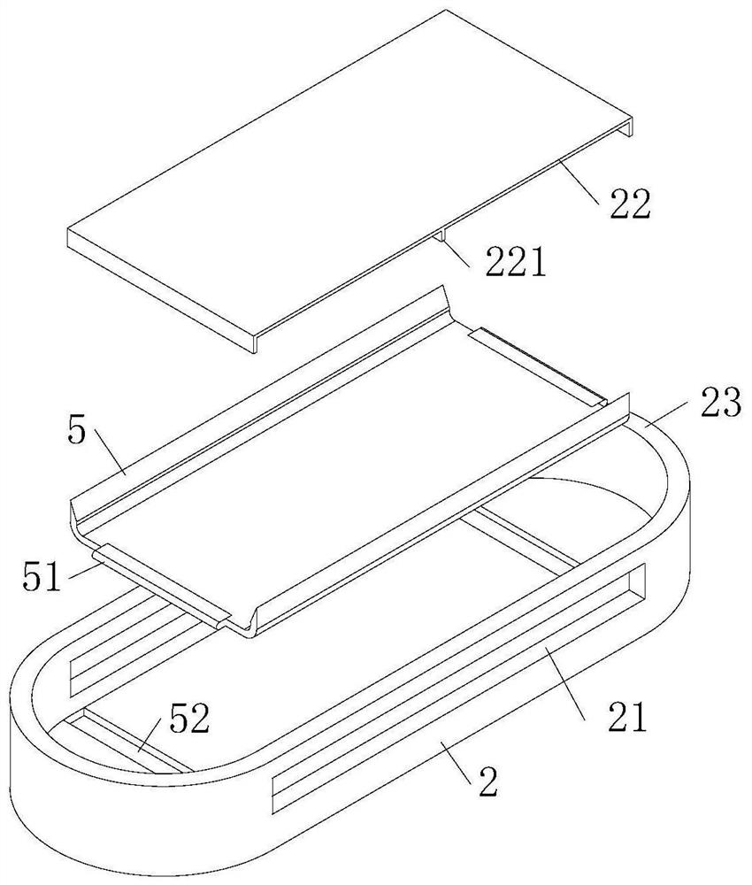
[0026] As an embodiment of the present invention, the bottom of the sampling vessel 1 is provided with a circumferentially distributed through groove 11, and a semiconductor refrigeration chip 4 is installed in the through groove 11; Above, the slicing disc 2 is at the same height position as the bottom surface of the sampling dish 1, and the slicing disc 2 seals the port of the through groove 11 in the sampling dish 1; Pathological slices are made by the slicing knife 22 and gradually fill up the space of the slicing disc 2, but there are still a large amount of unsliced ​​cells and tissues in the sampling dish 1, and the small space of the slicing disc 2 limits the production efficiency of the pathological slices; by setting The slicing disk 2 surrounding the sampling dish 1 makes the capacity of multiple slicing disks 2 meet the amount of cell tissue in the volume of the sampling dish 1, and enhances the number of pathological slices made at a time, and the through groove ar...


PUM


Abstract

The invention relates to the technical field of breast cancer detection, in particular to an auxiliary mammary duct in-situ cancer detection system which comprises a slice sampling device, a photographing unit, a storage module and an analysis module. The slice sampling device comprises a sampling vessel, a slice disc and a controller; due to the fact that IDC and DCIS have high similarity in thecellular morphology, the accuracy of distinguishing by directly utilizing a convolutional neural network algorithm from the cellular morphology is not high, and then the accuracy of DCIS detection isaffected. Therefore, through the slice disc arranged in the sampling vessel and the temperature control effect of the semiconductor chilling plate in the slice disc, the cell tissues in the sampling vessel are continuously made into pathological slices by the slicing knife in the slice groove, frozen and shaped in the slice disc to obtain a large number of pathological slices, and the pathologicalslices are provided for the analysis module for image recognition; and comprehensive analysis on pathological sections of cell tissues is achieved, so that the application effect of the auxiliary detection system for the breast duct in-situ cancer is improved.

Description

technical field [0001] The invention relates to the technical field of breast cancer detection, in particular to an auxiliary detection system for breast ductal carcinoma in situ. Background technique [0002] Ductal carcinoma in situ (DCIS) is the most common type of non-invasive breast cancer. The cancer cells are located in the breast ducts, have not yet penetrated the duct wall, and have not spread. Therefore, DCIS is not fatal, but DCIS is generally considered It is the precursor lesion of invasive ductal carcinoma (IDC), and DCIS may eventually develop into IDC without treatment; therefore, timely detection and treatment of DCIS become the key. [0003] At present, the pathological biopsy of breast ductal cells mainly relies on pathologists to observe pathological tissue samples under a microscope, and make a diagnosis based on empirical knowledge such as cell morphology, which heavily relies on the experience of pathologists; in recent years, image recognition methods...

Claims


Application Information

Patent Timeline
Patent Type & Authority Applications(China)
IPC IPC(8): G01N1/06G01N21/84G06K9/62G06N3/04G06N3/08G06T7/00
CPCG01N1/06G01N21/84G06T7/0012G06N3/08G06T2207/10061G06T2207/30024G06N3/045G06F18/214
Inventor 孙献甫
Owner HENAN CANCER HOSPITAL
Learn More