Pulmonary nodule automatic detection method and device and computer system

A technology for automatic detection of pulmonary nodules, applied in computer components, computing, image data processing, etc., can solve problems such as low sensitivity and excessive false positives, and achieve high detection sensitivity, noise reduction, and feature accuracy high effect

Pending Publication Date: 2021-01-05
SHANGHAI UNIV OF MEDICINE & HEALTH SCI +1
View PDF9 Cites 7 Cited by
  • Summary
  • Abstract
  • Description
  • Claims
  • Application Information

AI Technical Summary

Problems solved by technology

[0003] Judging from the current research status at home and abroad, many researchers are committed to the detection of pulmonary nodules, but the existing methods still have low sensitivity and too many false positives

Method used


Image

  • Pulmonary nodule automatic detection method and device and computer system
  • Pulmonary nodule automatic detection method and device and computer system
  • Pulmonary nodule automatic detection method and device and computer system

Examples


Embodiment 1

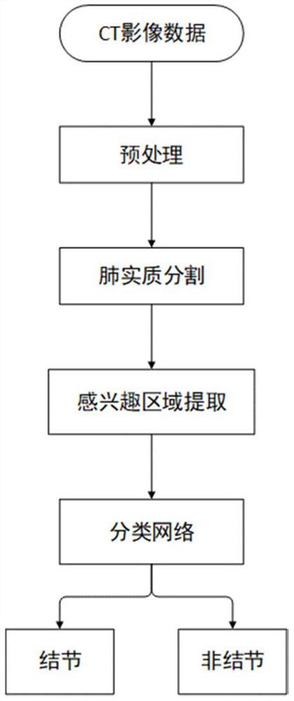
[0045] Such as figure 1 As shown, the present embodiment provides a method for automatic detection of pulmonary nodules, the method comprising:

[0046] Step 1, CT image data acquisition.

[0047] Step 2, sample preprocessing, including image filtering and window width and window level adjustment.

[0048] In this embodiment, the lung CT images are filtered by means of median filtering and mean filtering to reduce image noise and improve image quality, and then adjust the window width and window level of the lung CT images to enhance The contrast of the lung parenchyma area is obtained by enhancing the CT image sequence of the lung. Wherein, the specific operation of adjusting the window width and window level may be to set the pixels whose HU value is greater than 400 to 400, and to set the pixels whose HU value is less than -1000 to -1000.

[0049] Step 3, lung parenchyma segmentation.

[0050] The lung parenchyma segmentation mainly uses the "threshold method" to segmen...

Embodiment 2

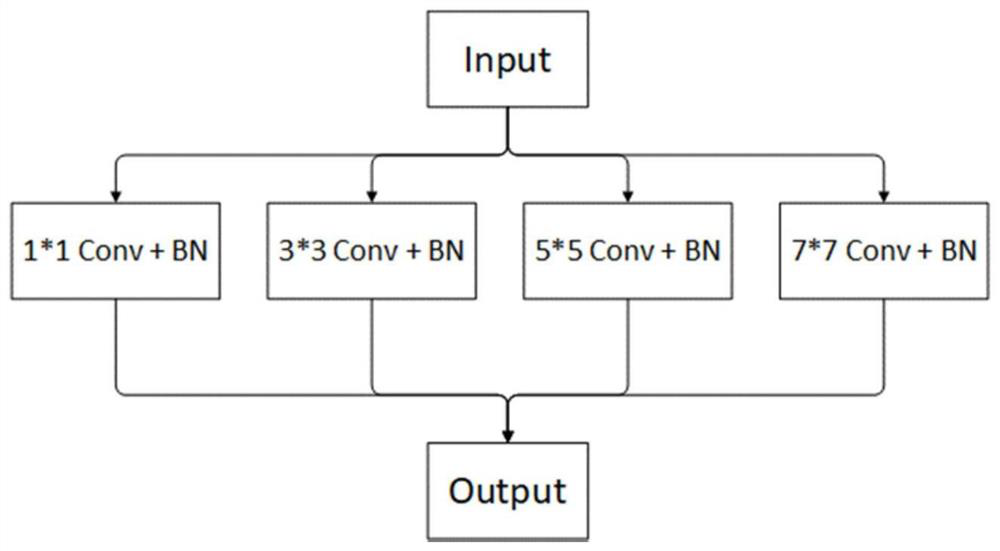
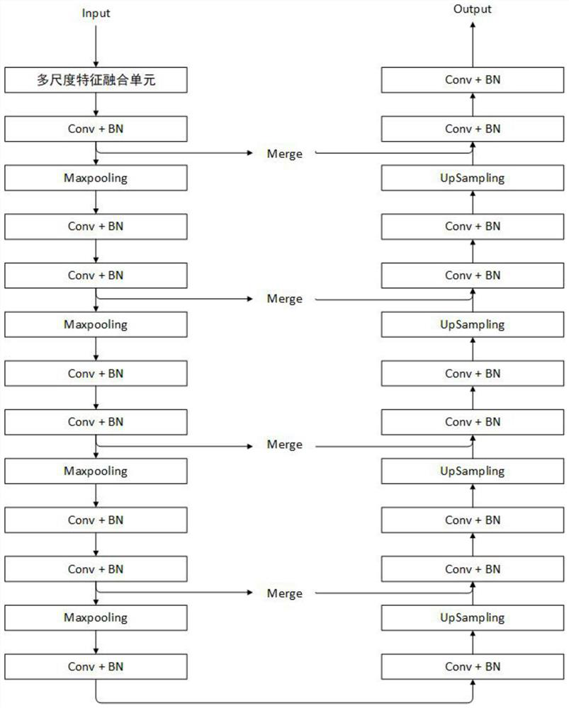
[0064] This embodiment provides an automatic detection device for pulmonary nodules, including: a CT image acquisition module, used to acquire a CT image to be detected; a preprocessing module, used to filter and enhance the CT image to be detected, to obtain lung enhancement The CT image sequence; the lung parenchyma segmentation module is used to segment the CT image sequence using the threshold method to obtain an image that only includes the lung parenchyma region; the region of interest extraction module is used to obtain the lung parenchyma segmentation module The image is cut into several image blocks, and the region of interest is obtained through a multi-scale feature fusion U-Net network model; the classification module is used to automatically detect and identify the region of interest using a 3D CNN model to obtain lung nodule detection result. All the other are with embodiment 1.

Embodiment 3

[0066] This embodiment provides a computer system for automatic detection of pulmonary nodules, including a processor and a memory storing processor-executable instructions; wherein, the processor is coupled to the memory, and is used to read program instructions stored in the memory , and in response, perform the steps in the method as described in Embodiment 1.


PUM


Abstract

The invention relates to a pulmonary nodule automatic detection method, a pulmonary nodule automatic detection device and a computer system. The method comprises the following steps: acquiring a CT image to be detected; performing filtering enhancement processing on the CT image to be detected to obtain a lung enhanced CT image sequence; segmenting the CT image sequence by adopting a threshold method to obtain an image only containing a pulmonary parenchyma region; cutting the image obtained by the pulmonary parenchyma segmentation module into a plurality of image blocks, and obtaining a region of interest through a multi-scale feature fusion UNet network model; and carrying out automatic detection and identification on the region of interest by adopting a 3D CNN model to obtain a pulmonary nodule detection result. Compared with the prior art, the method has the advantages of high detection sensitivity and precision and the like.

Description

technical field [0001] The invention relates to the field of computer aided detection, in particular to an automatic detection method, device and computer system for pulmonary nodules. Background technique [0002] Lung cancer is one of the tumor diseases with the highest mortality rate. Every year, more than 1.3 million people die from lung cancer in the world. In the early stage of lung cancer, its performance is not obvious, and more than 70% of lung cancer patients are basically in the advanced stage of lung cancer. According to relevant medical statistics, if lung cancer patients can get proper intervention treatment in the early stage of cancer, their 5-year survival rate can reach more than 90%, but the survival rate of lung cancer patients in stage 2-3 drops to 40%-5%. Therefore, "early detection, early diagnosis, and early treatment" is the key to improving the survival rate of lung cancer patients. Lung cancer usually manifests as pulmonary nodules in the early ...

Claims


Application Information

Patent Timeline
Patent Type & Authority Applications(China)
IPC IPC(8): G06T7/00G06T7/11G06T7/136G06K9/32G06K9/62G06N3/04
CPCG06T7/0012G06T7/11G06T7/136G06T2207/10081G06T2207/20081G06T2207/20084G06T2207/30064G06T2207/20132G06T2207/20021G06V10/25G06N3/045G06F18/253
Inventor 黄钢聂生东陈阳
Owner SHANGHAI UNIV OF MEDICINE & HEALTH SCI
Why Eureka
Try Eureka