3D co-culture model of fibroblasts and cancer cells as well as preparation method and application of model
A technology for fibroblasts and cancer cells, applied in the field of 3D co-culture model of directional migration of cancer cells and its preparation, can solve the problems that there is no mature model, and the growth process of individual cells cannot be observed separately in 3D co-culture models, to achieve research-friendly effects
- Summary
- Abstract
- Description
- Claims
- Application Information
AI Technical Summary
Problems solved by technology
Method used
Image

Examples
Embodiment 1
[0081] This embodiment provides a 3D co-culture system of fibroblasts and cancer cells.
[0082] (1) resuspend fibroblast cell line (BHK-21) and red fluorescent protein (RFP)-labeled cancer cell CaKi-1 in pre-cooled DMEM medium containing 5% serum;
[0083] (2) Add the thawed Matrigel to the fibroblast and cancer cell suspensions at 4°C, and the cell suspensions obtained after adding Matrigel meet the following conditions at the same time:
[0084] a. The number of cells is 5×10 5 cells / mL; b. The final concentration of Matrigel is 80%.
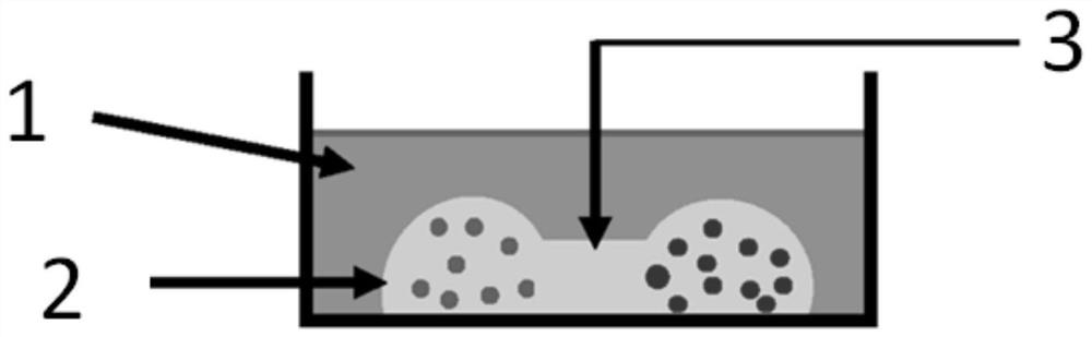
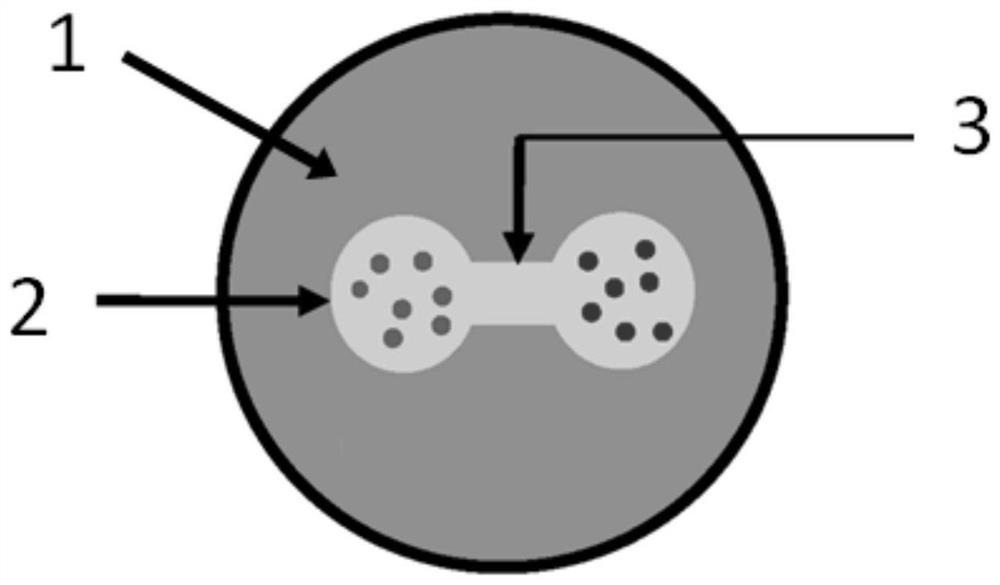
[0085] (3) Put 5 μL of fibroblast suspension mixed with Matrigel and 5 μL of cancer cell suspension adjacently in the same well of a 24-well culture plate, leaving an interval of about 2 mm between the two cell droplets;
[0086] (4) Place the culture plate in a 37°C incubator until the Matrigel suspension solidifies
[0087] (5) Construct Matrigel channel: drop liquid Matrigel (the liquid Matrigel is that Matrigel is diluted with DMEM to ...
Embodiment 2
[0093] This example is used to record the growth of cells in the 3D co-culture system obtained in Example 1.
[0094] For the convenience of comparison, in this example, the cells of fibroblasts and cancer cells (including CaKi-1 kidney cancer cells, HeLa cervical cancer cells, A375 human melanoma cells and A549 lung adenocarcinoma cells) were also observed when they were cultured alone. Morphological change.
[0095] (1) When fibroblasts are cultured alone, the obtained microscopic images are as follows: Figure 4 As shown, observing the morphological changes of fibroblasts on days 0, 3, 5, 8, 11, and 22 of culture shows that fibroblasts spontaneously form vasculature in 3D Matrigel (such as Figure 5 shown), and eventually shrink called spheroids.
[0096] (2) When cancer cells are cultured alone, the obtained microscopic images are as follows: Figure 6 As shown, cancer cells do not form fibroblast-like vasculature in 3D Matrigel suspension, but aggregate to form many sm...
Embodiment 3
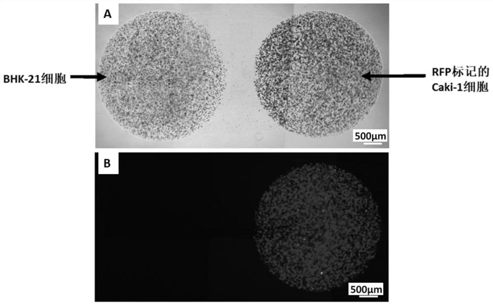
[0111] This example is used to study the affinity of different cancer cell lines to fibroblast vasculature in a 3D co-culture system. The construction method of the 3D co-culture system is the same as in Example 1.
[0112] The observed results are as Figure 13 As shown, microscopic views at high magnification fields show that different cancer cell lines (HeLa, A375 and A549) start to adhere to the vascular network formed by fibroblasts 4 days after inoculation.
[0113] In summary, through the co-culture model described in the present invention, it can be observed that after the fibroblasts come into contact with the cancer cells, the cancer cells begin to be attracted by the vessels of the fibroblasts; then the vascular network of the fibroblasts continues to The cancer cells move in one direction and at the same time begin to contract to form a spheroid and drag on the cancer cells attached to their vasculature; eventually, the vasculature of the fibroblast shrinks and ev...
PUM
 Abstract
Description
Claims
Application Information
 - R&D
- Intellectual Property
- Life Sciences
- Materials
- Tech Scout
- Unparalleled Data Quality
- Higher Quality Content
- 60% Fewer Hallucinations
Browse by: Latest US Patents, China's latest patents, Technical Efficacy Thesaurus, Application Domain, Technology Topic, Popular Technical Reports.
© 2025 PatSnap. All rights reserved.Legal|Privacy policy|Modern Slavery Act Transparency Statement|Sitemap|About US| Contact US: help@patsnap.com