Paranasal sinus tube stent and preparation method thereof

A technology for tube stents and sinus tubes, applied in the field of sinus tube stents and their preparation, can solve the problems of difficult to adjust the size in time, the degradation cycle is not synchronized with the tissue healing cycle, and the welding edge is broken.

Active Publication Date: 2020-08-14
INST OF MEDICAL DEVICES (SUZHOU) SOUTHEAST UNIV
View PDF8 Cites 5 Cited by
  • Summary
  • Abstract
  • Description
  • Claims
  • Application Information

AI Technical Summary

Problems solved by technology

[0003]The defect of the existing stent preparation technology is that: the nasal cavity of different individuals has different requirements for the stent size, and the stent prepared by the traditional method has low efficiency, poor repeatability and large size Difficult to adjust in time, the cost of using laser engraving to prepare side hollow brackets is high, and the wall thickness is not easy to control
For the preparation of tubular stents, the conventional stacking technology in the Z-axis direction has the following defects: (1) The preparation of stents with small diameters and complex pattern structures on the surface is difficult to achieve due to the limitation of instrument precision; (2) The stents prepared by stacking in the Z-axis direction The wall thickness needs to be thicker, otherwise it is difficult to achieve the requirement of high vertical stacking height; (3) Due to the limitation of wall thickness, the polymer material scaffold prepared by stacking in the Z-axis direction of 3D printing is hard and brittle and has almost no self-expanding performance
The stent wall produced by stacking 3D printing in the Z-axis direction is too thick and inelastic. The tubular 3D printing method has high requirements on the instrument itself and is not easy to obtain, and increases the cost of the stent. Limiting the implantation into the human body has a great medical risk, and the degradation cycle of the stent is not synchronized with the tissue healing cycle, the welding edge caused by hot gas welding is broken due to stress concentration, and key technical issues such as drug release rate and drug dose control waiting to be solved

Method used


Image

  • Paranasal sinus tube stent and preparation method thereof
  • Paranasal sinus tube stent and preparation method thereof
  • Paranasal sinus tube stent and preparation method thereof

Examples


preparation example Construction

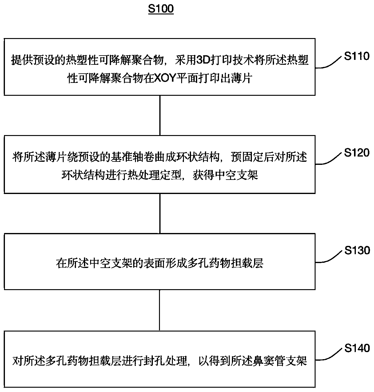
[0043] Such as figure 1 As shown, the first aspect of the present invention provides a method for preparing a sinus tube stent S100, which specifically includes the following steps S110-S140:

[0044] S110. Provide preset thermoplastic degradable polymers, and use 3D printing technology to print thin sheets of thermoplastic degradable polymers on the XOY plane.

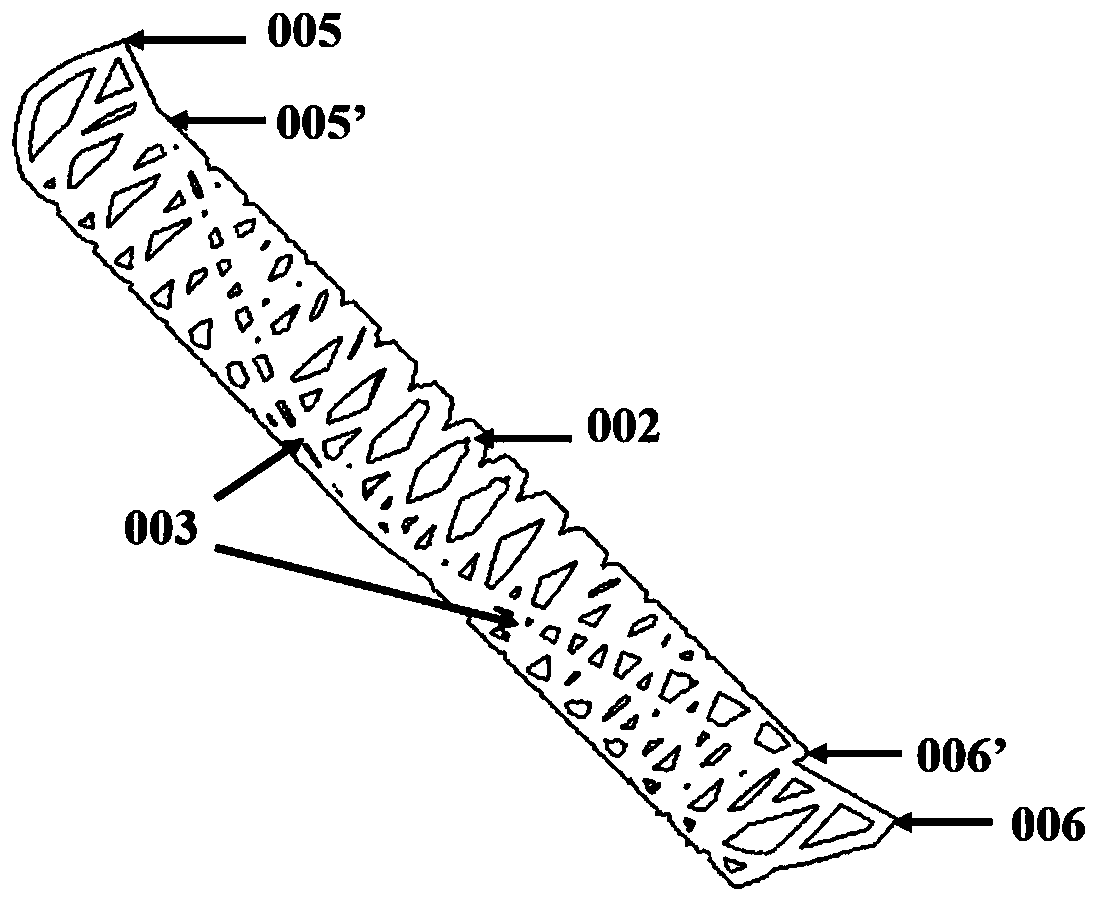
[0045] In view of the fact that the diameter of the stent prepared by the current 3D printing is not easy to change, and the wall thickness of the printed stent is too thick and inelastic, this embodiment uses a fused deposition 3D printer to print a thin sheet of thermoplastic degradable polymer on the XOY plane. The wall thickness is thinner and the diameter is controllable. Exemplary, combined with figure 2 As shown, in this embodiment, the sheet is configured as a parallelogram sheet, the sheet has a length ranging from 10 mm to 20 mm, a width ranging from 20 mm to 30 mm, and a thickness ranging from 0.1 mm to ...

Embodiment 1

[0070] The preparation method of sinus tube stent in this example comprises the following steps:

[0071] S1. Use 3D modeling software to build the side structure model of the bracket, and export the file format as .stl file, import it into the fused deposition 3D printer, and use polycaprolactone (Mv=40,000, PCL) as raw material to print patterns and shapes Unfold the structural sheet model for the sides of the parallelogram, i.e. print out the sheet. Wherein, the specific parameters of the side structure of the parallelogram sheet printed in this embodiment are as follows: length L=10.0 mm, width W=20 mm, sheet thickness t=0.1 mm.

[0072]S2, the parallelogram sheet obtained in step S1 according to the preset direction (refer to figure 2 The center line 004) is curled around a thin copper rod with a diameter of 3.0mm, and the two ends of the welding (reference figure 2 003) and pre-fixed with fine copper wire in the middle, put it in a vacuum drying oven at 40°C for the ...

Embodiment 2

[0079] The preparation method of sinus tube stent in this example comprises the following steps:

[0080] S1. Use 3D modeling software to build the side structure model of the bracket, and export the file format as .stl file, import it into the fused deposition 3D printer, and use polylactic acid glycolic acid (Mv=80,000, PLGA) as raw material to print patterns and shapes Unfold the structural sheet model for the sides of the parallelogram, i.e. print out the sheet. Wherein, the specific parameters of the side structure of the parallelogram sheet printed in this embodiment are as follows: length L=13.0 mm, width W=25.0 mm, sheet thickness t=0.2 mm.

[0081] S2, the parallelogram sheet obtained in step S1 according to the preset direction (refer to figure 2 The center line 004) is curled around a thin copper rod with a diameter of 4.0mm, and the two ends of the welding (reference figure 2 003) and pre-fixed with fine copper wire in the middle, put it in a vacuum drying oven...


PUM

PropertyMeasurementUnit
Viscosityaaaaaaaaaa
Lengthaaaaaaaaaa
Widthaaaaaaaaaa

Abstract

The invention provides a paranasal sinus tube stent and a preparation method thereof. The preparation method comprises the following steps: providing a preset thermoplastic degradable polymer, printing the thermoplastic degradable polymer into a thin sheet on an XOY plane by adopting a 3D printing technology, curling the thin sheet around a preset reference axis to form an annular structure, pre-fixing, and carrying out heat treatment shaping on the annular structure to obtain a hollow stent, and forming a porous drug carrying layer on the surface of the hollow stent, and carrying out hole sealing treatment on the porous drug carrying layer to obtain the sinus canal stent. The preparation method provided by the invention is not limited by the precision of a fused deposition type 3D printerinstrument, and ensures the consistency of forming conditions of all parts of the stent, so that the stent has good elasticity, the stent can be effectively prevented from escaping from a matrix in adeployment process, subsequent sudden release of drugs can be effectively prevented, and meanwhile, the problem of insufficient drug carrying capacity is also avoided, and thus the probability of medical accidents such as secondary blockage caused by the fact that granulation tissues of the nasal cavity grow into the space of the nasal cavity is prevented.

Description

technical field [0001] The invention belongs to the technical field of biomedical devices, and in particular relates to a sinus tube bracket and a preparation method thereof. Background technique [0002] Nasal obstruction is a common clinical disease. It often leads to poor breathing of patients, and even causes facial mutilation and paralysis, which directly affects people's normal life. At present, the main method of interventional therapy is to use the catheter delivery system to send the drug-eluting stent on the surface into the blocked nasal cavity, which can not only expand the narrow nasal cavity, but also use the drug to achieve the repair function. [0003] The disadvantages of the existing stent preparation technology are: the nasal cavity of different individuals has different requirements for the stent size, the stent prepared by the traditional method is low in efficiency, the repeatability is poor, and the size is difficult to adjust in time. Higher, the wa...

Claims


Application Information

Patent Timeline
IPC IPC(8): A61F2/90B33Y70/00B33Y80/00
CPCA61F2/90B33Y80/00B33Y70/00A61F2310/0097A61F2240/001A61F2210/0004A61F2210/0014
Inventor 白晶王先丽程兆俊邵怡薛烽储成林汪丰
Owner INST OF MEDICAL DEVICES (SUZHOU) SOUTHEAST UNIV
Why Eureka
Try Eureka