Construction method of rare earth up-conversion nano-probe labeled virus
A rare earth up-conversion and nano-probe technology, applied in the field of biotechnology and materials, can solve the problems of unfavorable long-term tracing, poor photostability, high background noise, etc., and achieve the effect of high scientific research application prospects
- Summary
- Abstract
- Description
- Claims
- Application Information
AI Technical Summary
Problems solved by technology
Method used
Image
Examples
Embodiment 1
[0030] Preparation of envelope biotinylated influenza virus
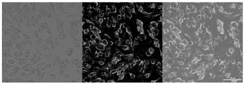
[0031] MDCK cells were cultured in a cell culture dish with a diameter of 10 cm, DSPE-PEG2000-Biotin reagent (final concentration 1 μg / ml) was added to the medium, and the culture dish was placed in a 37°C carbon dioxide incubator for 18-24h. When the cell density in the culture dish grows to 80-90%, discard the medium, wash the cells twice with serum-free medium to remove residual serum, add 5ml serum-free medium and 10μl influenza virus concentrate, mix well and place Incubate in a carbon dioxide incubator at 37°C for 2 hours, shaking the culture dish every half hour. After incubation, the supernatant was discarded, and the complete medium containing 2% FBS and DSPE-PEG2000-Biotin was added. Place the culture dish in a 37°C carbon dioxide incubator for 48-72h. When 50% of the cells died, the cells and supernatant were collected in a centrifuge tube, and the cells were completely broken up by repeated freezing an...
Embodiment 2
[0033] Preparation of envelope biotinylated influenza virus
[0034] MDCK cells were cultured in a cell culture dish with a diameter of 10 cm, DSPE-PEG2000-Biotin reagent (final concentration 5 μg / ml) was added to the medium, and the culture dish was placed in a 37°C carbon dioxide incubator for 18-24h. When the cell density in the culture dish grows to 80-90%, discard the medium, wash the cells twice with serum-free medium to remove residual serum, add 5ml serum-free medium and 10μl influenza virus concentrate, mix well and place Incubate in a carbon dioxide incubator at 37°C for 2 hours, shaking the culture dish every half hour. After incubation, the supernatant was discarded, and the complete medium containing 2% FBS and DSPE-PEG2000-Biotin was added. Place the culture dish in a 37°C carbon dioxide incubator for 48-72h. When 50% of the cells died, the cells and supernatant were collected in a centrifuge tube, and the cells were completely broken up by repeated freezing an...
Embodiment 3
[0036] Preparation of envelope biotinylated influenza virus
[0037] MDCK cells were cultured in a cell culture dish with a diameter of 10 cm, DSPE-PEG2000-Biotin reagent (final concentration: 50 μg / ml) was added to the culture medium, and the culture dish was placed in a 37°C carbon dioxide incubator for 18-24h. When the cell density in the culture dish grows to 80-90%, discard the medium, wash the cells twice with serum-free medium to remove residual serum, add 5ml serum-free medium and 10μl influenza virus concentrate, mix well and place Incubate in a carbon dioxide incubator at 37°C for 2 hours, shaking the culture dish every half hour. After incubation, the supernatant was discarded, and the complete medium containing 2% FBS and DSPE-PEG2000-Biotin was added. Place the culture dish in a 37°C carbon dioxide incubator for 48-72h. When 50% of the cells died, the cells and supernatant were collected in a centrifuge tube, and the cells were completely broken up by repeated f...
PUM
 Abstract
Description
Claims
Application Information
 - R&D
- Intellectual Property
- Life Sciences
- Materials
- Tech Scout
- Unparalleled Data Quality
- Higher Quality Content
- 60% Fewer Hallucinations
Browse by: Latest US Patents, China's latest patents, Technical Efficacy Thesaurus, Application Domain, Technology Topic, Popular Technical Reports.
© 2025 PatSnap. All rights reserved.Legal|Privacy policy|Modern Slavery Act Transparency Statement|Sitemap|About US| Contact US: help@patsnap.com


