
Antibody biological activity detection method based on surface plasma resonance technology

A technology of surface plasmon and antibody biology, applied in measuring devices, instruments, and material analysis through optical means, can solve the problem of large variability, long detection cycle, and inability to meet the needs of rapid detection of in vitro activity of antibodies targeting immune checkpoints and other problems to achieve the effect of simple steps, high efficiency and easy operation

Active Publication Date: 2020-06-30
江苏华泰疫苗工程技术研究有限公司
View PDF3 Cites 0 Cited by
  • Summary
  • Abstract
  • Description
  • Claims
  • Application Information

AI Technical Summary

Problems solved by technology

However, none of the existing detection methods can meet the needs of rapid detection of in vitro activity of antibodies targeting immune checkpoints, for example, mixed lymphocyte reaction (Mixed lymphocyte reaction, MLR) and antigen-specific stimulation assays (antigen-specific recall assays ) can detect biological activity by detecting the secretion of IFN-γ, but the detection cycle is longer and takes 4-5 days
Moreover, these methods use primary cells. Due to the differences brought about by different individuals and the complexity of experimental operations, the variability of these two detection methods is very large.
In addition, in vitro activity detection methods based on T cell proliferation, cytotoxicity, etc. also require the use of primary cells and complicated operations, which are not satisfactory as a rapid detection method for the biological activity of immune checkpoint antibodies.

Method used


Image

  • Antibody biological activity detection method based on surface plasma resonance technology
  • Antibody biological activity detection method based on surface plasma resonance technology
  • Antibody biological activity detection method based on surface plasma resonance technology

Examples


Embodiment Construction

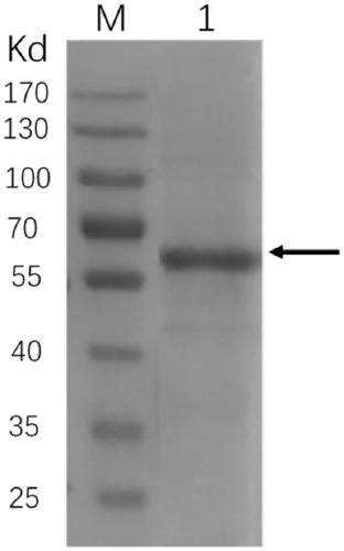
[0020] Expression and Purification of Human LAG3 Extracellular Domain Protein

[0021] Construction of the recombinant expression plasmid of LAG3 extracellular domain protein: Using the cDNA of human LAG3 extracellular domain protein as a template, the gene fragment of LAG3 extracellular domain protein was amplified by PCR and cloned into the expression vector pTT5.

[0022] Expression and purification: The human LAG3 extracellular region protein recombinant vector constructed above was transfected into HEK293 cells for transient protein expression. The expressed cell supernatant was collected, separated and purified with a nickel ion affinity column (GE company product) and molecular exclusion chromatography column Superdex 200 (GE company product), and the obtained human LAG3 extracellular domain protein was used for subsequent detection method analysis.

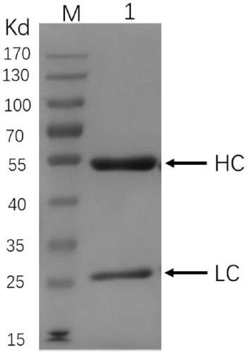
[0023] Expression and purification of anti-LAG3 monoclonal antibody relatlimab

[0024] Construction of anti-LAG3 mono...


PUM


Abstract

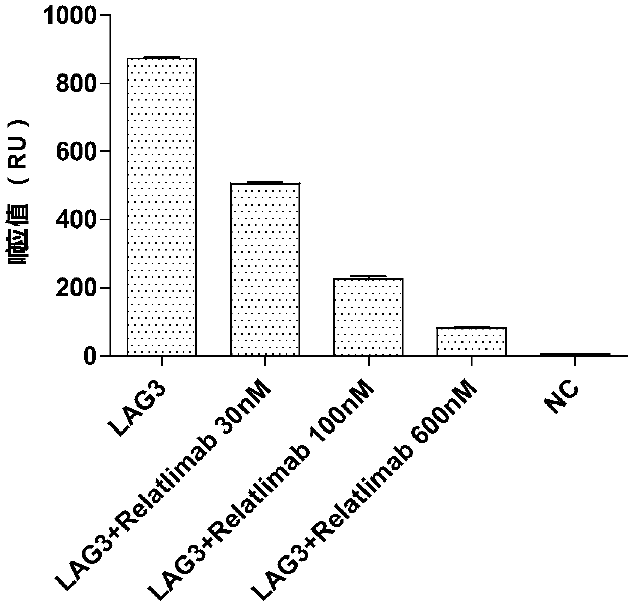
The invention provides an antibody biological activity detection method based on a surface plasma resonance technology, and particularly relates to the technical field of biological detection, human Burkitt's lymphoma cell strain Daudi cells highly expressed by an LAG3 receptor are selected to be combined with a C1 chip, and the combined cells are fixed on the surface of the chip; when the LAG3 iscombined with cells on the surface of the chip, a response signal is captured through a biomacromolecule interaction instrument Biacore T200; when the anti-LAG3 antibody is added, the combination ofLAG3 and cells is reduced, the response value is reduced, and the antibody activity at the cellular level is realized by comparing the response value so as to realize rapid detection. The method is simple in step, easy to operate, capable of achieving real-time dynamic monitoring and high in efficiency, has a specific recognition effect, and has great advantages in antibody drug screening, batch release, stability monitoring, even process development and other applications of immune checkpoint targets.

Description

technical field [0001] The invention belongs to the technical field of biological detection, and in particular relates to an antibody biological activity detection method based on surface plasmon resonance technology. Background technique [0002] Immune checkpoint blocking antibodies can fight cancer by enhancing the function of the immune system, and this type of cancer immunotherapy has become one of the important methods in the treatment of various malignant tumors in recent years. Immune checkpoints can be stimulatory or inhibitory, with co-stimulatory proteins signaling to promote an immune response to pathogens, and inhibitory in the opposite direction. Among them, the inhibitory molecules cytotoxic T lymphocyte-associated antigen 4 (CTLA-4) and programmed cell death receptor 1 (PD-1) are the two most studied immune checkpoint molecules. CTLA-4 is the first immune checkpoint target for clinical use, and its inhibitor ipilimumab was approved for marketing in 2011. Pr...

Claims


Application Information

Patent Timeline
IPC IPC(8): G01N33/569G01N21/552
CPCG01N33/56966G01N21/553Y02A50/30
Inventor 雷高新叶华跃王永智乔正邱文娜陈凌彭福中吴宏斌
Owner 江苏华泰疫苗工程技术研究有限公司
Learn More