
Fluid control device and method for enriching and coloring cells

A fluid control device and cell technology, which can be used in measurement devices, sample preparation for testing, material analysis by optical means, etc., and can solve the effects of loss, inability to quantify, capture efficiency, dosage, repeated cleaning, non-specific adsorption, etc. To achieve the effect of high detection rate, easy operation, improved enrichment degree and diagnostic sensitivity

Pending Publication Date: 2019-12-20
JINAN UNIVERSITY
View PDF0 Cites 1 Cited by
  • Summary
  • Abstract
  • Description
  • Claims
  • Application Information

AI Technical Summary

Problems solved by technology

Traditional cell enrichment methods include centrifugal enrichment method, biochemical capture method, membrane filtration enrichment method, etc. Although centrifugation method can enrich cells, the volume, shape and weight of cells vary, making some cells Discarded; biochemical capture methods usually use biomolecules or chemical molecules that can bind to cell surface molecules such as antibodies, ligands, polar or non-polar chemical molecules attached to a solid phase, and then capture cells by affinity to achieve enrichment However, the capture efficiency is affected by intermolecular affinity, molecular dosage, repeated cleaning, non-specific adsorption, etc., so losses will also occur; the membrane filtration enrichment method is to filter through a filter membrane with a smaller pore size than the cells to be captured, and the cells are completely Retained by the filter membrane on the surface of the membrane, it is an ideal enrichment method, but because the filter pressure is difficult to control, some cells adhere tightly to the membrane and cannot be eluted, and some cells will also be lost
[0003] Staining the cells allows medical staff and researchers to obtain the test results of the required cells from the microscopic examination. Commonly used staining methods for cells in body fluid samples include bacterial dye staining, fluorescent antibody combined with cell staining, etc., but the commonly used staining methods It is greatly affected by human factors; the staining process requires repeated washing of the slides, and some cells that are not firmly fixed will be washed away; the dripping of coloring solution and elution time depend on the experience of the operator, which is random and cannot be quantified. Difficult to repeat; open handling exposes operators to hazards

Method used


Image

  • Fluid control device and method for enriching and coloring cells
  • Fluid control device and method for enriching and coloring cells
  • Fluid control device and method for enriching and coloring cells

Examples


Embodiment 1

[0038] In this embodiment, the fluid control device of the present invention is used to perform Gram staining to color bacteria. The microporous filter membrane used in the fluid control device is a track-etched polyethylene terephthalate (PETTE) membrane with a pore size of 0.45 μm. The components of the pre-filled solution 1-9 are pre-filled with the components of the Gram staining solution. The following are ammonium oxalate crystal violet solution, ultrapure water, iodine solution, ultrapure water, 95% alcohol, ultrapure water, ultrapure water, saffron red solution, and ultrapure water.

[0039] 1. Sample collection and processing

[0040] (1) The patient coughs up about 5mL of sputum and spits it into a sterile 50mL centrifuge tube.

[0041] (2) Add 5 mL of 4% NaOH solution, shake for 1 minute, and let stand for 15 minutes.

[0042] (3) Add 0.067mol / L phosphate buffered saline (PBS) to 50mL, and centrifuge at 3000xg for 20 minutes.

[0043] (4) Pour off the supernatant...

Embodiment 2

[0059] In this embodiment, the fluid control device of the present invention is used to carry out the acid-fast staining method to color the acid-fast bacilli. The filter membrane used in the fluid control device is a track-etched polyethylene terephthalate (PETTE) membrane with a pore size of 0.45 μm. The components of the acid-fast staining solution are pre-filled in chambers 1-9 of the pre-filled solution, followed by stone Fuchsin carbonate solution, ultrapure water, ultrapure water, acidic alcohol, ultrapure water, ultrapure water, methylene blue solution, ultrapure water, ultrapure water.

[0060] 1. Sample collection and processing

[0061] (1) The patient coughs up about 5mL of sputum and spits it into a sterile 50mL centrifuge tube.

[0062] (2) Add 5 mL of 4% NaOH solution, shake for 1 minute, and let stand for 15 minutes.

[0063] (3) Add 0.067mol / L phosphate buffered saline (PBS) to 50mL, and centrifuge at 3000xg for 20 minutes.

[0064] (4) Pour off the superna...

Embodiment 3

[0080]In this embodiment, the fluid control device of the present invention is used to perform auramine O fluorescent staining to make acid-fast bacilli stain with fluorescence. The filter membrane used in the fluid control device is a track-etched polyethylene terephthalate (PETTE) membrane with a pore size of 0.45 μm. The components of the acid-fast staining solution are pre-filled in chambers 1-9 of the pre-filled solution, followed by gold Amine O dye solution, ultrapure water, ultrapure water, acid alcohol, ultrapure water, ultrapure water, potassium permanganate solution, ultrapure water, ultrapure water.

[0081] 1. Sample collection and processing

[0082] (1) The patient coughs up about 5mL of sputum and spits it into a sterile 50mL centrifuge tube.

[0083] (2) Add 5 mL of 4% NaOH solution, shake for 1 minute, and let stand for 15 minutes.

[0084] (3) Add 0.067mol / L phosphate buffered saline (PBS) to 50mL, and centrifuge at 3000xg for 20 minutes.

[0085] (4) Pou...


PUM

PropertyMeasurementUnit
pore sizeaaaaaaaaaa

Abstract

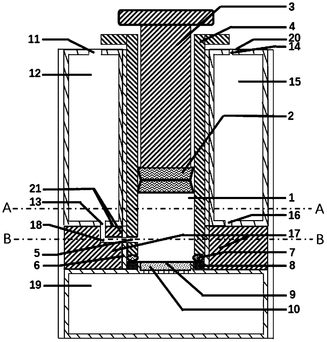
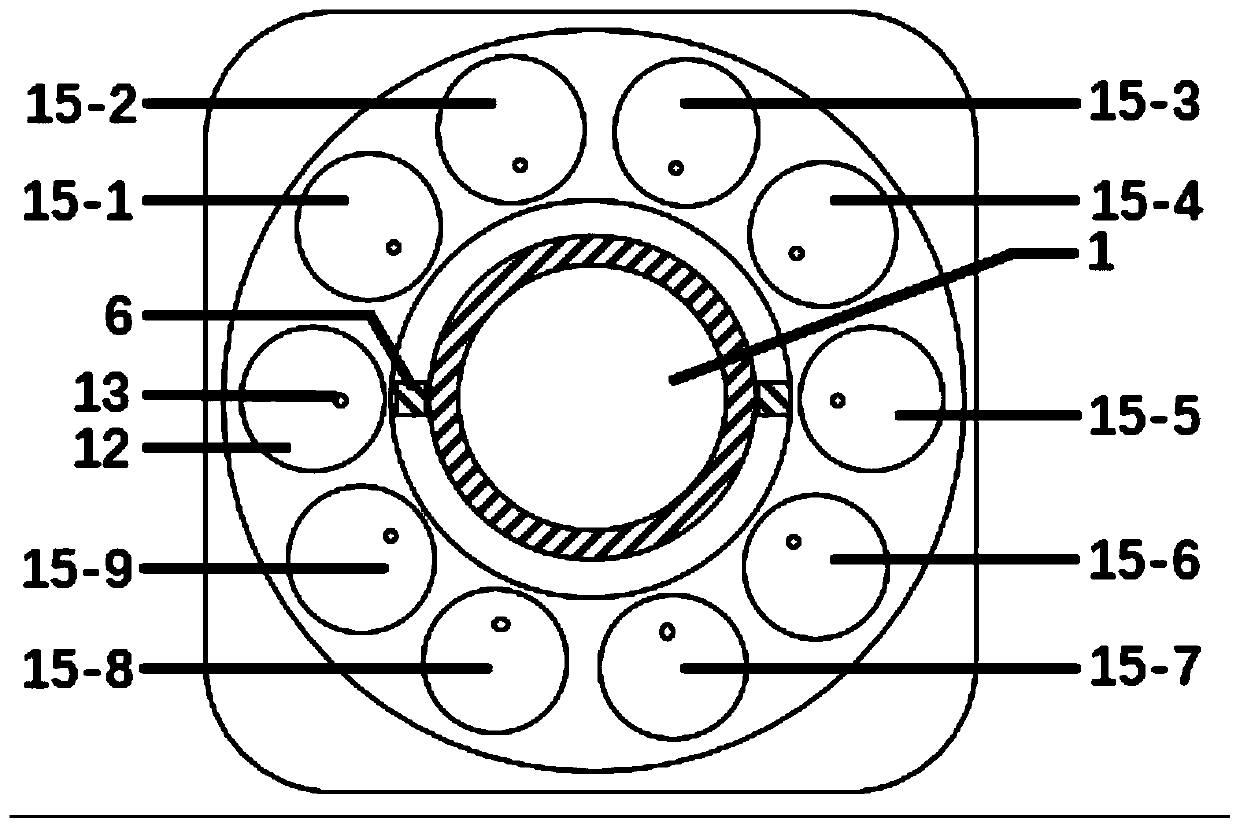
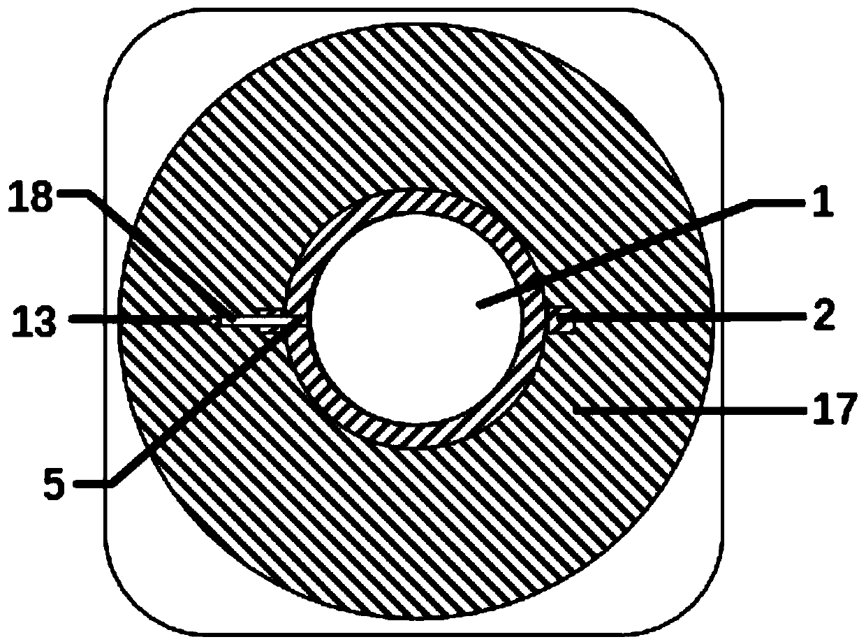
The invention discloses a fluid control device and method for enriching and coloring cells. The device comprises a filtering cavity body, a rotary disc, a sample chamber, liquid pre-loading chambers and a waste liquid chamber, the filtering cavity body is detachably arranged on the rotary disc, the sample chamber and the plurality of liquid pre-loading chambers are uniformly arranged in the circumferential direction of the filtering cavity body, the waste liquid chamber is arranged at the bottom of the rotary disc. The method generates pressure difference by pushing and pulling the piston, rotates the filtering cavity to form a channel to flow fluid into the filtering cavity and carry out filtering, and enriches the filtered cells on the microporous filter membrane and colors the cells onthe microporous filter membrane. The invention quantitatively adds liquid in the coloring process, reduces the randomness, improves the repeatability, and can avoid the risk caused by the repeated contact of operators with samples. The invention relates to the technical field of medical sample processing.

Description

technical field [0001] The invention relates to the technical field of medical sample processing, in particular to a fluid control device and method for enriching and coloring cells. Background technique [0002] Making slices of prokaryotic or eukaryotic cells in body fluid samples and observing them under a microscope is a common method for diagnosing diseases in medical tests, and is also an important means in biological research. The cells in the sample are enriched, stained and other processed. Traditional cell enrichment methods include centrifugal enrichment method, biochemical capture method, membrane filtration enrichment method, etc. Although centrifugation method can enrich cells, the volume, shape and weight of cells vary, making some cells Discarded; biochemical capture methods usually use biomolecules or chemical molecules that can bind to cell surface molecules such as antibodies, ligands, polar or non-polar chemical molecules attached to a solid phase, and t...

Claims


Application Information

Patent Timeline
Patent Type & Authority Applications(China)
IPC IPC(8): G01N21/84G01N21/64G01N1/34G01N1/30
CPCG01N21/84G01N21/6458G01N1/34G01N1/30
Inventor 唐勇
Owner JINAN UNIVERSITY
Learn More