
Cervix uteri and vagina histocyte sample acquisition device for HPV detection experiment

A tissue cell and sample collection technology, applied in colposcopy, application, inoculation and ovulation diagnosis, etc., can solve problems such as patient injury, patient vaginal lining injury, delaying the best time for treatment, etc., to avoid cross infection, convenient operation, avoid damage effect

Active Publication Date: 2019-07-23
AFFILIATED HOSPITAL OF ZUNYI MEDICAL COLLEGE
View PDF9 Cites 9 Cited by
  • Summary
  • Abstract
  • Description
  • Claims
  • Application Information

AI Technical Summary

Problems solved by technology

[0004] When the existing cervicovaginal tissue cell collection device is inserted into the patient's vagina, most of the duckbill tablets are used when dilating the patient's vagina, which is easy to cause damage to the inner wall of the patient's vagina; the depth of the device's entry cannot be controlled, and it is easy to cause harm to the patient; Clearly photograph the inner wall of the cervix and vagina of the patient, so that the patient cannot detect the condition in time and delay the best time for treatment

Method used


Image

  • Cervix uteri and vagina histocyte sample acquisition device for HPV detection experiment
  • Cervix uteri and vagina histocyte sample acquisition device for HPV detection experiment
  • Cervix uteri and vagina histocyte sample acquisition device for HPV detection experiment

Examples


Embodiment

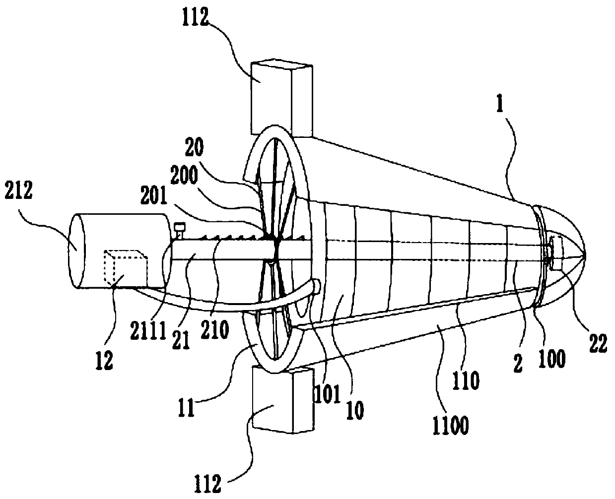
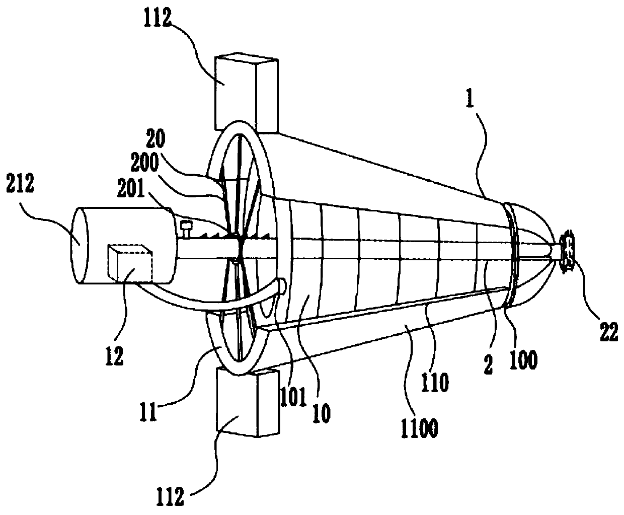
[0024] Example: such as figure 1 , 2 A cervicovaginal tissue cell sample collection device for HPV detection experiments mainly includes a vaginal expansion element 1, a collection element 2, and a power supply device; the vaginal expansion element 1 includes a round platform vaginal expansion air bag 10, a support buckle plate 11, and a micro air pump 12, The round platform Yin expansion air bag 10 is a hollow structure, such as Figure 7 As shown, and the smaller end of the round platform Yin expansion airbag 10 is provided with a connecting ring 100, and the larger end is provided with an inflation port 101, and is symmetrically arranged on the outer sidewall of the round platform Yin expansion airbag 10, and the two supporting gussets 11 are The arc-shaped gusset matching the shape of the outer wall of the round platform Yin-expanding airbag 10, the radian of the supporting gusset 11 is 1 / 3-2 / 3π, and the micro air pump 12 is connected with the Round table Yin-expanding ai...


PUM


Abstract

The invention discloses a cervix uteri and vagina histocyte sample acquisition device for an HPV detection experiment, and belongs to the technical field of medical apparatuses. The cervix uteri and vagina histocyte sample acquisition device mainly comprises a cervix uteri extending element, an acquisition element and a power source device, wherein the acquisition element comprises limiting fixingframes, an acquisition adjusting rod and an acquisition head. According to the cervix uteri and vagina histocyte sample acquisition device disclosed by the invention, the vagina of a patient is expanded through the cervix uteri extending element, so that the acquisition adjusting rod can conveniently enter for histocyte acquisition, and shooting of the cervix uteri and the vagina inner wall of the patient, by a camera is clearer; when being in contact with the vaginal orifice of the patient, the cervix uteri and vagina histocyte sample acquisition device is limited by the acquisition adjusting rod, so that the situation that the patient is damaged because a cervix uteri extending air sac and supporting pinch plates are inserted too deep is avoided; through a replaceable banister brush layer on the acquisition head, histocytes in the cervix uteri and vagina of the patient are sampled, so that cross infection is avoided; shooting is performed through the micro camera on the acquisitionhead, so that the illness state of the cervix uteri and the vagina is convenient for the patient to know, the disease can be found out timely, and treating can be performed as soon as possible; and the cervix uteri and vagina histocyte sample acquisition device disclosed by the invention is simple in structure, safe and high-efficiency, convenient to operate and suitable for wide popularization.

Description

technical field [0001] The invention belongs to the technical field of medical devices, and in particular relates to a device for collecting cervicovaginal tissue cell samples for HPV detection experiments. Background technique [0002] HPV is a kind of papillomavacuolating virus A belonging to Papovaviridae. It is a spherical DNA virus that can cause squamous epithelial proliferation of human skin and mucous membranes, manifested as common warts, genital warts and other symptoms. With the rapid increase in the incidence of genital warts in sexually transmitted diseases and the increase in cervical cancer and anal cancer, HPV infection has attracted more and more attention. [0003] HPV detection is a means to deal with human papillomavirus. It is clinically proven that the virus is a DNA virus. Humans are the only host of HPV. It will cause disease. At present, scientists have proved that the HPV virus transmitted through sexual life is the main cause of cervical cancer. ...

Claims


Application Information

Patent Timeline
IPC IPC(8): A61B10/02A61B1/303A61B1/04
CPCA61B1/04A61B1/303A61B10/02A61B2010/0216
Inventor 陈祖翼肖代敏黄健李琼瑶闵迅陈泽慧鄢仁晴
Owner AFFILIATED HOSPITAL OF ZUNYI MEDICAL COLLEGE
Learn More