Method for testing proliferative response of drug to tumor cell derived from patient

A tumor cell, proliferation response technology, applied in the field of tumor cell proliferation response, can solve problems such as low usability, poor reproducibility and stability, complex amplification or reduction of components, and insufficiently characterized samples, etc., to achieve predictive The effect of the reaction

Pending Publication Date: 2019-07-19
SHANGHAI BIODURO BIOLOGICS
View PDF5 Cites 1 Cited by
  • Summary
  • Abstract
  • Description
  • Claims
  • Application Information

AI Technical Summary

Problems solved by technology

[0003] However, current 3D cell culture technologies have certain limitations, including poor reproducibility and stability, complexity of components, difficulty in scaling up or down, and / or the need for improved physiological substrates
Specifically, anti-proliferation predictive assays for oncology drug candidates tested in a 3D growth environment using patient-derived tumor cells suffer from high heterogeneity, low proliferation, and / or low availability of well-characterized samples, produce unpredictable results, therefore, there is an urgent need in the art to overcome such limitations

Method used


Image

  • Method for testing proliferative response of drug to tumor cell derived from patient
  • Method for testing proliferative response of drug to tumor cell derived from patient
  • Method for testing proliferative response of drug to tumor cell derived from patient

Examples


Embodiment 1

[0033] In Vitro Proliferation Assay

[0034] Thaw patient-derived tumor cells in a 37°C water bath until the frozen cell suspension is fully thawed to form a slurry solution, slowly transfer the cell suspension to a 50ml conical tube through a cell strainer, and pour into the conical tube Fill up with conditioned medium warmed to 37°C; spin the conical tube at 800 RPM for 5 min at 4°C to pellet the cells; remove the supernatant carefully without disturbing the cell pellet by aspirating the supernatant, Resuspend the cell pellet in 1-2 ml of warmed conditioned medium, count the cells using TC10, and finally remove 10 μl of the cell suspension and transfer to a tube for trypan blue exclusion assay to assess cell density and vial Overall health of cells, counting live-dead cells.

[0035] use (ECM from Trevigen) to coat a 6-well plate, dilute the ECM to 1:20 with ice-cold conditioned medium without growth factors and supplements, add 1 ml of diluted Cultrex solution to each we...

Embodiment 2

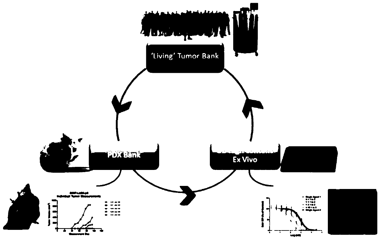
[0048] Use of Patient-Derived Xenografts (PDX)

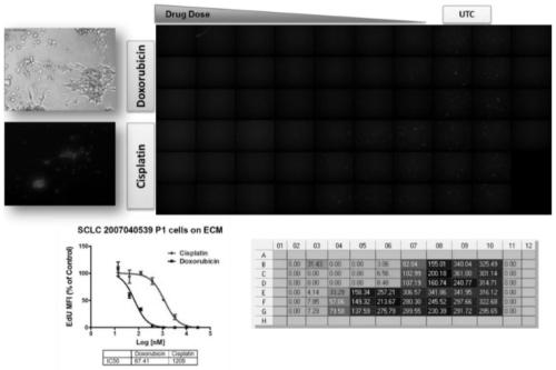
[0049] Fresh or banked patient-derived tumor cells were injected into immunocompromised mice, cells were harvested and plated to form spheroids, and spheroids were processed ex vivo with chemotherapeutic candidates, cisplatin, and doxorubicin Drug treatment, cell proliferation measured by HC imaging of EdU incorporation assay. ( Figure 1A -B)

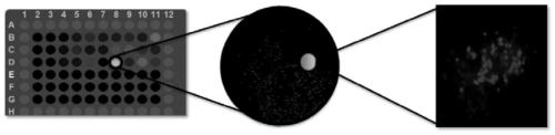
[0050] Cell images are captured and processed using a variety of algorithms corresponding to the experiment. Select DAPI on channel 1 to identify valid objects and EdU on channel 2 to identify DNA-incorporated cell populations, record and integrate the intensity of each pixel for each channel to obtain the total intensity, divide the total intensity In total pixels, the mean fluorescence intensity (MFI) of the channel was obtained. The integrated mean fluorescence intensity of multiple cells was divided by the total number of cells in each well to obtain the mean value of MFI. To norma...


PUM


Abstract

The invention relates to use of a 3D cell culture technology in an in-vitro drug test method, and more particularly to a method for testing a proliferative response of a drug to tumor cell derived from a patient, which can reliably screen the candidate drugs by predictive proliferation assay for efficacy and / or mechanism of action. The method comprises the steps of: (1) obtaining cells from a biopsy or tumor resection material; (2) culturing the obtained cells on a 3D extracellular matrix; (3) treating the tumor cells cultured on the 3D extracellular matrix with drug treatment; (4) performinghigh-content imaging on the processed tumor cells; and (5) evaluating the high-content imaging of the processed tumor cells, and testing the proliferative response of the drug to the tumor cells derived from the patient.

Description

technical field [0001] The present invention relates to the use of 3D cell culture technology in ex vivo drug testing methods, in particular to a method of testing the proliferative response of drugs to patient-derived tumor cells. Background technique [0002] The current trend in drug development and disease-related research is to gradually move away from the use of non-human primate models. Recently, in NIH research, the use of chimpanzees has been significantly reduced. The method allows cells to acquire a natural 3D phenotype and can enhance cell proliferation, differentiation and function, while traditional 2D (two-dimensional) cell culture methods are known to be unable to accurately represent the real 3D world of disease progression, drug testing and / or biochemistry and physiology studies. As a result, advanced technologies based on in vitro platforms have been and will continue to be developed, utilizing complex scaffolds and extracellular matrices to support cell...

Claims


Application Information

Patent Timeline
IPC IPC(8): C12Q1/02
CPCG01N33/5011C12N2503/02G01N2500/10
Inventor 赛勒斯·库罗斯·米尔赛迪汤马斯·本杰明·布鲁迪胡庸
Owner SHANGHAI BIODURO BIOLOGICS
Why Eureka
Try Eureka