A New Method for Determining the Integrity of Type I Collagen Triple Helix Structure
A technology for structural integrity and collagen, which is applied in the field of judging the integrity of type I collagen triple helix, can solve the problems of collagen triple helix structure damage and affect biological activity, etc., to achieve quantitative analysis, good reproducibility, Simple operation effect
- Summary
- Abstract
- Description
- Claims
- Application Information
AI Technical Summary
Problems solved by technology
Method used
Image
Examples
Embodiment 1
[0072] (1) Take the collagen sample of aquatic carp skin to be tested, add pre-cooled 0.5M acetic acid solution, dissolve it at 10°C and divide it into tubes A and B, and add an appropriate amount of pepsin;
[0073] (2) Tube A is always in a water bath at 10°C, and tube B is placed in a water bath at 40°C;
[0074] (3) At the set time, draw samples from tubes A and B into tubes A1 and B1 respectively;
[0075] (4) Place tubes A1 and B1 in an ice water bath quickly, and add pepsin inhibitor (Pepstatin);
[0076] (5) Repeat the operation from steps (3) to (4) and change the set time. A1 and B1 are obtained at 0 minutes, A2 and B2 at 30 minutes, A3 and B3 at 1 hour, and A4 and B4 at 2 hours , get A5, B5 at 3h, A6, B6 at 5h;
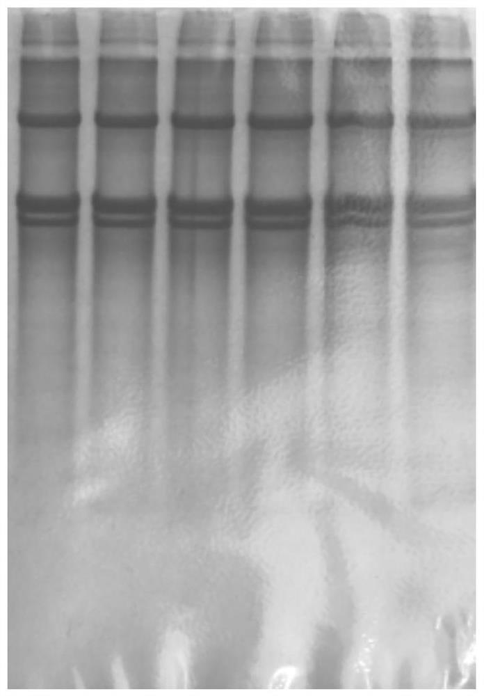
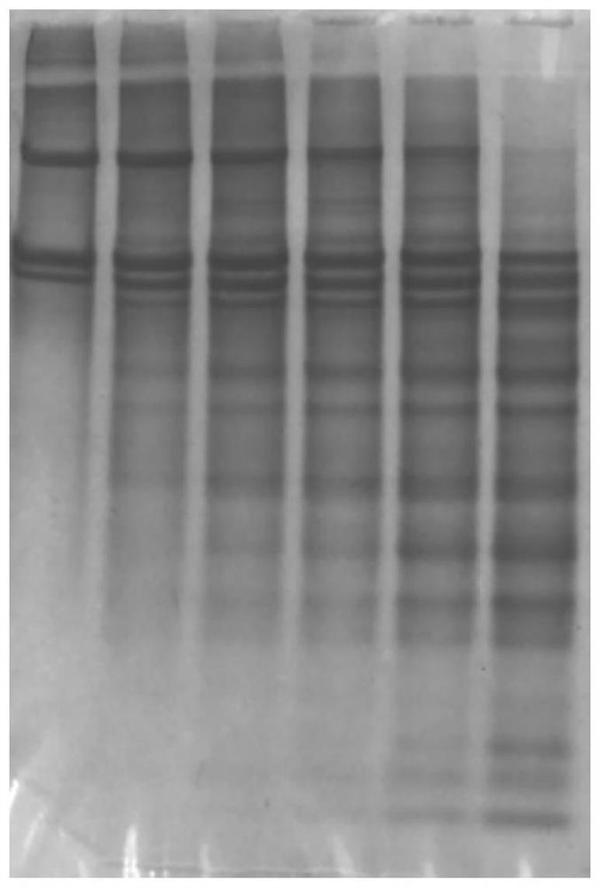
[0077] (6) Prepare electrophoresis samples from the obtained A1-6 and B1-6 samples, such as figure 1 and 2 as shown, figure 1 The 6 bands shown in are respectively A1, A2, A3, A4, A5, A6, figure 2 The six bands shown in are B1, B2, B3, B4, B5, and B6...
Embodiment 2
[0083] (1) Take the collagen-rich animal tissue pigskin collagen sample to be tested, add pre-cooled 0.1M acetic acid solution, dissolve at 5°C and divide into tubes A and B, and add appropriate amount of pepsin;
[0084] (2) Tube A is always in a water bath at 0°C, and tube B is placed in a water bath at 80°C;
[0085] (3) At the set time, draw samples from tubes A and B into tubes A1 and B1 respectively;
[0086](4) Place tubes A1 and B1 in an ice water bath quickly, and add pepsin inhibitor (Pepstatin);
[0087] (5) Repeat the operation from steps (3) to (4) and change the set time. A1 and B1 are obtained at 0 minutes, A2 and B2 at 30 minutes, A3 and B3 at 1 hour, and A4 and B4 at 2 hours , get A5, B5 at 3h, A6, B6 at 5h;
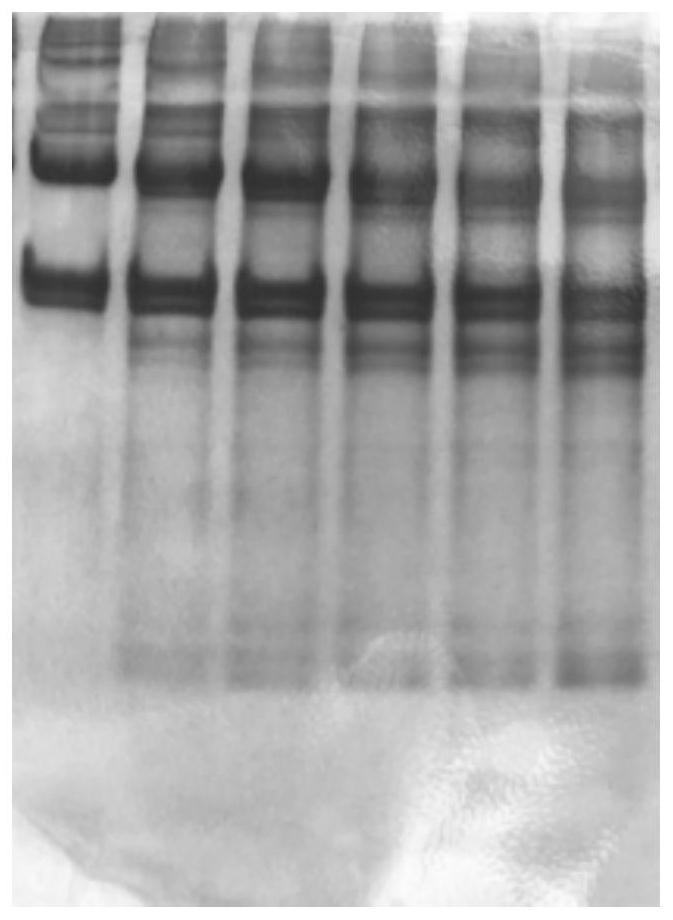
[0088] (6) Prepare electrophoresis samples from the obtained A1-6 and B1-6 samples; image 3 and 4 as shown, image 3 The 6 bands shown in are respectively A1, A2, A3, A4, A5, A6, Figure 4 The 6 bands shown in are B1, B2, B3, B4, B5, B6 respective...
Embodiment 3
[0094] (1) Take the beef tendon collagen sample of animal tissue rich in collagen to be tested, add pre-cooled 0.5M acetic acid solution, dissolve at 0°C, and divide into tubes A and B, and add appropriate amount of pepsin;
[0095] (2) Tube A is always in a water bath at 10°C, and tube B is placed in a water bath at 70°C;
[0096] (3) At the set time, draw samples from tubes A and B into tubes A1 and B1 respectively;
[0097] (4) Place tubes A1 and B1 in an ice water bath quickly, and add pepsin inhibitor (Pepstatin);
[0098] (5) Repeat the operation from steps (3) to (4) and change the set time. A1 and B1 are obtained at 0 minutes, A2 and B2 at 30 minutes, A3 and B3 at 1 hour, and A4 and B4 at 2 hours , get A5, B5 at 3h, A6, B6 at 5h;
[0099] (6) Prepare electrophoresis samples from the obtained A1-6 and B1-6 samples; Figure 5 and 6 as shown, image 3 The 6 bands shown in are respectively A1, A2, A3, A4, A5, A6, Figure 4 The 6 bands shown in are B1, B2, B3, B4, B5,...
PUM
 Abstract
Description
Claims
Application Information
 - R&D
- Intellectual Property
- Life Sciences
- Materials
- Tech Scout
- Unparalleled Data Quality
- Higher Quality Content
- 60% Fewer Hallucinations
Browse by: Latest US Patents, China's latest patents, Technical Efficacy Thesaurus, Application Domain, Technology Topic, Popular Technical Reports.
© 2025 PatSnap. All rights reserved.Legal|Privacy policy|Modern Slavery Act Transparency Statement|Sitemap|About US| Contact US: help@patsnap.com


