
Method and system for mass segmentation in mammogram

A line image and mass technology, applied in the field of machine learning and digital medical image processing and analysis, can solve problems such as relying on manual design, and achieve the effects of improving accuracy, improving precision, and expanding the number of

Active Publication Date: 2020-12-29
SOUTH CENTRAL UNIVERSITY FOR NATIONALITIES
View PDF9 Cites 0 Cited by
  • Summary
  • Abstract
  • Description
  • Claims
  • Application Information

AI Technical Summary

Problems solved by technology

However, all these traditional large-scale segmentation methods heavily rely on hand-crafted features

Method used


Image

  • Method and system for mass segmentation in mammogram
  • Method and system for mass segmentation in mammogram
  • Method and system for mass segmentation in mammogram

Examples


no. 1 example

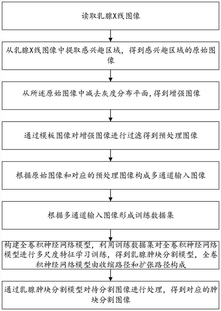
[0037] The first embodiment of the present invention, such as figure 1 As shown, a method for mass segmentation in mammography images, including:

[0038] Read mammogram images;

[0039] Extract the region of interest from the mammogram image to obtain the original image of the region of interest;

[0040] Subtract the gray distribution plane from the original image to obtain the enhanced image;

[0041] Filtering the enhanced image through the template image to obtain a preprocessed image;

[0042] Construct a multi-channel input image according to the original image and the corresponding preprocessed image;

[0043] Form a training dataset from multi-channel input images;

[0044] Construct a fully convolutional neural network model, use the training data set to perform multi-scale feature learning and training on the fully convolutional neural network model, and obtain a breast mass segmentation model. The fully convolutional neural network model consists of a contracti...


PUM


Abstract

The invention provides a method and a system for segmenting lumps in a mammary gland X-ray image, and the method comprises the steps: reading the mammary gland X-ray image, and extracting a region ofinterest from the mammary gland X-ray image to obtain a corresponding original image; Subtracting the gray level distribution trend plane from the original image to obtain an enhanced image; Filteringthe enhanced image through the template image to obtain a preprocessed image; Forming a multi-channel input image according to the original image and the preprocessed image; Forming a training data set according to the multi-channel input image; Constructing a full convolutional neural network model, and performing multi-scale feature learning training on the full convolutional neural network model by using the training data set to obtain a breast mass segmentation model; And processing the to-be-segmented image through the mammary gland lump segmentation model to obtain a corresponding lumpsegmentation image. According to the method, the original image and the preprocessed image with the remarkable lump appearance are input through multiple channels, and then multi-scale processing is carried out, so that the lump segmentation image with higher precision is obtained.

Description

technical field [0001] The invention relates to the fields of machine learning and digital medical image processing and analysis, in particular to a method and system for segmenting masses in mammogram images. Background technique [0002] In the treatment of breast cancer, early diagnosis and early treatment are considered to be the main methods to improve the survival rate of breast cancer. Since mammography is one of the standard techniques for early detection and diagnosis of breast cancer, automatic segmentation of masses in mammographic images is crucial for further quantitative and qualitative analysis. However, since a mammogram is a two-dimensional projection image, it is difficult to clearly identify a mass when the surrounding breast structure is similar to the intensity distribution of the mass, and the mass may have irregular shapes, low contrast, and different sizes, so Mass segmentation in radiographs remains quite challenging. [0003] There are many relate...

Claims


Application Information

Patent Timeline
Patent Type & Authority Patents(China)
IPC IPC(8): G06T7/11G06T7/12G06N3/04G06N3/08
Inventor 徐胜舟
Owner SOUTH CENTRAL UNIVERSITY FOR NATIONALITIES
Learn More