Glutamine optical probe and preparation method and application thereof
An optical probe, glutamine technology, applied in the field of optical probes, can solve in situ, real-time, dynamic, high-throughput and high spatio-temporal resolution detection of non-viable cells and subcellular organelles, not suitable for living cell research, Separation, extraction, purification and other issues, to achieve the effect of eliminating sample processing steps, large dynamic changes in fluorescence, and easy maturation
- Summary
- Abstract
- Description
- Claims
- Application Information
AI Technical Summary
Problems solved by technology
Method used
Image


Examples
preparation example Construction
[0072] The present invention also provides a preparation method for the above-mentioned glutamine optical probe, comprising the following steps: 1) incorporating the nucleic acid sequence encoding the glutamine optical probe described herein into an expression vector; 2) transferring the expression vector into a host cell 2) cultivating the host cell under conditions suitable for the expression of the expression vector, 3) isolating the glutamine optical probe.
[0073] The term "nucleic acid" or "nucleotide" as used in the present invention may be in the form of DNA or RNA. Forms of DNA include cDNA, genomic DNA or synthetic DNA. DNA can be single-stranded or double-stranded. DNA can be either the coding strand or the non-coding strand. The term "variant" as used herein in reference to a nucleic acid may be a naturally occurring allelic variant or a non-naturally occurring variant. These nucleotide variants include degenerate variants, substitution variants, deletion varia...
Embodiment 1
[0158] Example 1: Plasmids expressing glutamine-binding proteins
[0159] The glutamine-binding protein QBP (GlnH) gene in the Agrobacterium agrobacterium gene was amplified by PCR, and the PCR product was recovered after gel electrophoresis and digested with BamHI and EcoRI, and the pRSETb vector was subjected to corresponding double digestion. After ligation with T4 DNA ligase, the product was used to transform MachI, and the transformed MachI was spread on LB plates (ampicillin 100ug / mL), and cultured at 37°C overnight. After plasmid extraction of the growing MachI transformants, PCR identification was performed. After the positive plasmid is correctly sequenced, the subsequent plasmid construction is carried out.
Embodiment 2
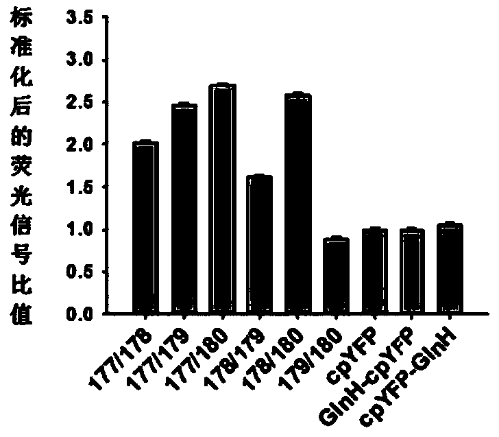
[0160] Example 2: Expression and detection of cpYFP optical probes at different fusion sites
[0161] In this example, the following sites were selected for fusion with cpYFP based on pRSETb-QBP to obtain the corresponding pRSETb-QBP-cpYFP plasmid: 177 / 178, 177 / 179, 177 / 180, 178 / 179, 178 / 180 or 179 / 180.
[0162] The DNA fragment of cpYFP was generated by PCR, and the DNA fragment was inactivated after adding phosphorous to the 5' end, and the pRSETb-glutamine binding protein linearized vector containing different break sites was amplified by reverse PCR at the same time. The linearized pRSETb-QBP and the phosphorylated cpYFP fragment at the 5' end were ligated under the action of PEG4000 and T4 DNA ligase to produce recombinant plasmids. These plates were picked on the Kodak multifunctional in vivo imaging system and picked up under the excitation of the FITC channel. Yellow The fluorescent clones were sequenced by Beijing Liuhe Huada Gene Technology Co., Ltd. Shanghai Branc...
PUM
 Abstract
Description
Claims
Application Information
 - R&D
- Intellectual Property
- Life Sciences
- Materials
- Tech Scout
- Unparalleled Data Quality
- Higher Quality Content
- 60% Fewer Hallucinations
Browse by: Latest US Patents, China's latest patents, Technical Efficacy Thesaurus, Application Domain, Technology Topic, Popular Technical Reports.
© 2025 PatSnap. All rights reserved.Legal|Privacy policy|Modern Slavery Act Transparency Statement|Sitemap|About US| Contact US: help@patsnap.com